


PEER REVIEWED
Understanding Geographic Atrophy in Advanced
Non-Exudative Age-Related Macular Degeneration:
a Teaching Case Report
James Rogala, OD, FAAO
Abstract
Age-related macular degeneration (AMD) continues to be a common cause of debilitating vision loss for many older Americans. While the exudative form of the disease is treatable, no effective treatment exists for the non-exudative form. This case report illustrates the clinical presentation of geographic atrophy in advanced non-exudative AMD, demonstrates how optometrists may assist these individuals, and discusses potential future prevention and treatment options.
Key Words: non-exudative age-related macular degeneration, exudative age-related macular degeneration, geographic atrophy, disciform scarring, choroidal neovascularization, anti-vascular endothelial growth factor
Background
Age-related macular degeneration (AMD) is a common condition which, as the name implies, results in acquired degenerative changes involving the macular region of the eye. The initial presentation is typically the focal deposition of abnormal amounts of extracellular debris, known as drusen, within and around Bruch’s membrane, the acellular layer separating the retinal pigment epithelium (RPE) from the choriocapillaris. As drusen accumulate, the normal exchange of nutrients and metabolic waste products between photoreceptors and their underlying vascular supply may become impaired, resulting in gradually progressive bilateral vision dysfunction. Eventually, RPE cell death may ensue and lead to focal atrophy of overlying photoreceptors and the underlying choriocapillaris. The resultant sharply demarcated atrophic patches at the posterior pole are referred to as geographic atrophy (GA). These atrophic areas usually first appear in the perifoveal region and may slowly expand to include the fovea itself, producing severe central vision loss due to what is considered the end-stage of “non-exudative” AMD. In some instances, choroidal neovascularization (CNV) may develop and be accompanied by leakage and the accumulation of extracellular fluid, which often causes abrupt vision loss due to what is termed “exudative” AMD.1,2
AMD is a multifactorial disorder with a multitude of genetic, environmental, and nutritional components contributing to its development. The main risk factor by far is age, and the condition is almost by definition non-existent in those under the age of 50.3 Demographically, Caucasians are affected more often than those of other ethnicities. Positive family history and certain genetic profiles also predispose individuals to the development of AMD. The major modifiable risk factor is cigarette smoking.4 AMD has consistently been found to be a leading cause of severe irreversible vision loss in developed countries throughout the world.5,6,7 Unfortunately, there is currently no treatment for the non-exudative form, which constitutes up to 90% of all cases of AMD.1,2 Nonetheless, optometrists can benefit these patients by reducing their risk of progression, detecting treatable exudative manifestations, and providing vision rehabilitation. Clinical trials, which may enable treatment in the near future, are under way.
The following case report is intended to help facilitate discussion and understanding of key concepts related to the clinical presentation and management of GA amongst optometry students, residents and practitioners.
Case Description
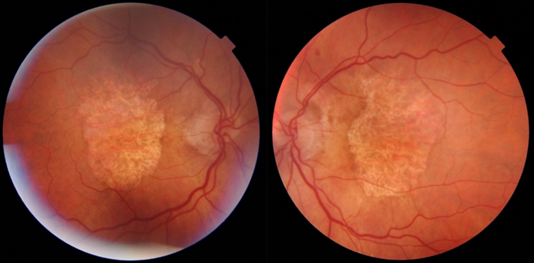
Figure 1. Central geographic atrophy OD and OS.
Click to enlarge
A 91-year-old Caucasian gentleman presented for examination as advised by his primary care physician. He was accompanied by his daughter who provided a history of long-standing vison loss, described 5 years earlier as “legal blindness” due to “macular degeneration,” with no recent changes. She also reported that he had undergone cataract surgery on both eyes several years ago and that a metallic foreign body was removed from his left eye many years ago. His most recent eye exam had been “a few” years prior with an ophthalmologist. The daughter further explained that her father had not seen the ophthalmologist since then because his other health concerns had taken precedence and his vision had not seemed to change. Family ocular health was uncertain, but it sounded as if he may have had siblings (brother and sister) who also experienced vison loss from macular degeneration in their later years. His daughter reported that he was being treated for “heart disease,” diabetes, hypertension and high-cholesterol, and that he had undergone multiple cardiac bypass surgeries. A list of current medications was obtained that was consistent with and appropriate for the conditions described. The patient was being visited weekly at his home by a nurse practitioner to ensure adequate management of the aforementioned cardiovascular conditions, with periodic office visits to the primary care provider who had advised ocular examination.

Table 1. Click to enlarge
Best-corrected visual acuity was 20/200 in each eye at distance and near with a prescription of OD: +0.50 -0.50 x 100, OS: +0.75 -0.75 x 10⁵ using a +3.50 near add. Due to difficulty with fixation, cover test results were undeterminable, but extraocular muscles were unrestricted. Color vision testing using Ishihara plates was unreliable. Pupils were equal, round and reactive to light with no relative afferent pupillary defect. Confrontation visual fields were full to finger counting OU, while Amsler grid testing revealed central scotomas in each eye. Intraocular pressures were measured as OD: 13 mmHg, OS: 14 mmHg via non-contact tonometry after applanation tonometry was deemed unsafe/impractical due to excessive head movement and blinking. Slit lamp examination revealed normal lids and lashes, quiet conjunctiva, and corneas that were clear centrally with mild arcus OU. There was no significant staining of conjunctiva or cornea with fluorescein in either eye, and both anterior chambers were deep and quiet with clear, well-positioned posterior chamber intraocular lenses in each eye. Von Herrick estimation of both anterior chamber angles was 4/4. The patient’s pupils were dilated with 1% tropicamide OU. Posterior segment evaluation revealed well-circumscribed areas of chorioretinal atrophy centrally in both eyes (Figure 1). Optic nerves were well-perfused with minimal cupping, distinct margins and temporal peripapillary atrophy OU. Retinal vasculature was essentially normal apart from trace dot and blot hemorrhages with very mild arteriole narrowing and arteriovenous crossing changes at the posterior pole of each eye (Table 1).
A diagnosis of advanced non-exudative AMD resulting in central GA was made based on the history obtained from the patient’s daughter and the distinct fundus appearance at presentation. Given the stable nature of the patient’s condition and lack of any currently proven treatment to reverse his vision loss, the patient and his daughter were reassured that he would not completely lose sight from this disorder and informed of the potential for low vision services to maximize his remaining vision. Following this discussion, both the patient and his daughter expressed a desire to pursue a low vision consultation as suggested, and appropriate referral was made in-house to a trusted optometric colleague with residency training and experience in low vision rehabilitation. In addition, they were advised to continue home monitoring with the Amsler grid and to return for a comprehensive eye examination in 12 months, or sooner if any changes were noticed. In discussion with the patient’s daughter, more frequent follow-up was deemed unnecessary at this time given the apparent stability of his condition and the difficulties of traveling to the clinic in addition to attending to his other healthcare needs.
Summary of low vision consultation
The patient was seen a month later for a comprehensive vision rehabilitation evaluation. Our low vision specialist noted that he was hoping for help with reading his bible, bird-watching and gardening. There were no significant changes in history, medications or findings from the comprehensive eye examination conducted a month previously. A tritan color vision defect was detected with D-15 testing (tested OU) and a moderate decline in contrast sensitivity OU was elicited using the Mars letter chart. The patient and his daughter were provided with extensive counseling on the nature and extent of his vision loss and numerous vision aid devices were trialed. From among those demonstrated, the Eschenbach Magno 8x Binoculars were found to be especially useful and information was provided on how to obtain a loaner with the potential for purchase. He was also provided with a completed application for the state’s audiobook library. Finally, they were informed that he qualified for legal blindness status and they were referred to the appropriate state agency to obtain services for independent living. Prior to being discharged, they were advised to contact low vision services if they had problems using any of the devices or services offered by them and to follow up with me as directed.
Education Guidelines
The target audience for this case report as a learning exercise would primarily be optometry students or residents. Specifically, this case could serve as a teaching/learning tool within a didactic course on posterior segment disease because the focus is on medical management and the disease process. It would not be appropriate for a low vision course (though a case report on AMD emphasizing that aspect of care certainly could be).
This particular case could also be presented in a clinical grand rounds format to supplement direct patient care activities as part of a clinical rotation or externship. In fact, during the early months of the COVID-19 pandemic we used cases such as this for that very purpose while our students were prohibited from participating in direct patient care. While this case was not used in that manner, those that were effectively bridged the gap in patient care activities during the months that was not possible.
A final way in which this type of case report can be integrated into the curriculum is as a remediation or make-up assignment. For example, I am considering employing it in this manner within our posterior segment disease course when examination results indicate an individual requires additional instruction (outside the class as a whole) to achieve an acceptable level of competency on this topic. In this role, the student could be provided with the case report as a self-directed learning exercise. After allowing sufficient time for review, assessment might entail having the student answer the discussion questions presented below. In this context, the literature suggests this would perhaps best assess critical thinking and clinical decision-making via an oral or short-answer examination (as opposed to multiple-choice questioning). 8,9
Learning objectives
After reviewing this case report and subsequent discussion, one should be able to:
- Appreciate the burden imposed by AMD and GA on society and individual patients
- Understand the pathophysiology behind AMD and GA
- Differentiate AMD and GA from similar-appearing disorders
- Understand the utility of various ancillary tests and when each is indicated
- Appropriately counsel and manage patients presenting with AMD and GA
Key concepts
- The epidemiology, demographics and morbidity associated with AMD and GA
- The underlying pathophysiology of AMD and GA
- The differential diagnoses of AMD and GA
- The utility of ancillary testing in the diagnosis and management of AMD and GA
- Proper management of patients with various manifestations of AMD and GA
Discussion questions
1. Discuss how problematic AMD and GA are on both a population and individual level:
- what is the incidence/prevalence?
- what are the main risk factors, in terms of age, ethnicity and environmental influences?
- what are the consequences in terms of vision, overall well-being, cost?
2. Discuss the pathophysiology of AMD and GA:
- which anatomical structures are involved and how does this alter ocular function?
- what detrimental changes occur at the biochemical, cellular and histological level?
- what is the typical progression of non-exudative AMD from onset through end stage?
3. Discuss how optometrists diagnose AMD and GA:
- what is the relationship between drusen and AMD?
- what other disorders display drusen and/or chorioretinal atrophy?
- how can an optometrist distinguish between AMD/GA and conditions that look similar?
4. Discuss the clinical tools available to help diagnose and manage AMD and GA:
- what different types of imaging facilitate diagnosis and management? How?
- what non-imaging tests (labs, etc.) are useful? How?
- how is progression monitored in and out of the office?
5. Discuss the current standard of care and what the future holds for managing AMD and GA:
- what is the appropriate optometric management of each stage of AMD?
- what is the role of nutrition supplementation at each stage of AMD?
- when should an optometrist refer a patient with AMD? To whom? For what?
Teaching and assessment methodology
The purpose of this case report is to provide a real-world example of AMD with GA that can be used as a platform for discussion to increase understanding and improve patient care. Educators may choose to present only the case findings initially and provide the discussion questions as homework, to be completed individually or in groups, reserving further discussion for a subsequent meeting. Alternatively, the entire case report, including the discussion section, could be assigned as a self-directed individual or group study assignment.
Assessment may be formative (reviewing discussion question answers together with feedback) or summative (to evaluate proficiency). For the latter, the instructor might wish to create multiple-choice or short-answer questions based on the discussion questions provided. In either case, the goal should be to ensure competency, critical thinking and the ability to apply the concepts discussed to diverse clinical scenarios.
Discussion
Age-related macular degeneration remains the most common cause of severe irreversible vision loss in developed countries such as the United States and Canada.10,11 It is conventionally designated as being either the “dry” non-exudative form or the “wet” exudative form. While the advent of anti-vascular endothelial growth factor (anti-VEGF) medications has enabled treatment and, for some, reversal of vision loss in those with exudative AMD, there is currently no proven treatment for the more common non-exudative form of AMD.12 This is significant because more than a million Americans are afflicted with GA.13
Visual impairment due to AMD is a substantial societal burden. Various epidemiological studies have found the non-exudative manifestation of the disorder, for which there is no current treatment, to account for approximately 90% of all AMD cases.1 Unfortunately, demographic trends mean the incidence of both forms of AMD is increasing and expected to rise dramatically in the coming years. Within the United States, the number of individuals with AMD increased from 1.75 million to 2.07 million between 2000 and 2010, an increase of 18%, and that number is projected to more than double to 5.44 million by 2050.14 In terms of healthcare costs, the World Health Organization estimated the direct costs of vision impairment due to AMD in the United States, Canada and Cuba alone (WHO subregion AMR-A) at nearly $100 billion (in 2008 USD).15 Thus, given the magnitude of the problem, it is imperative that practicing optometrists understand this disorder and keep abreast of the latest developments in diagnosis and management in order to properly care for this large and growing patient population.
While the pathogenesis of AMD is complex and multifactorial, certain risk factors have been recognized. Age is the most significant, with those over age 75 being three times more likely to develop the disorder compared to their cohorts age 65-74.6,7 Genetic factors are another important risk factor and probably help account for the fact that AMD is more prevalent in Whites than in Blacks while prevalence in the Hispanic and Chinese populations falls somewhere in between.16,17 Specific genes have been shown to either predispose or be protective for AMD development, most notably the complement factor H (CFH) gene.1-4 Several environmental and lifestyle factors have also been investigated. Smoking has been identified as the main modifiable risk, roughly tripling the likelihood of developing AMD compared to non-smokers and increasing the risk of progression in those with AMD.3,18,19 Moreover, the combination of genetic and environmental risk factors, such as smoking, appears to have a synergistic effect on the progression of AMD.20 It is also likely that obesity, poor diet and cardiovascular disorders such as hypertension are risk factors though to what extent remains unclear.21-23 It is less certain whether other proposed factors, such as UV exposure, pose a significant risk.24
The complex pathogenesis of AMD means there is still much to be learned and a detailed discussion is well beyond the scope of this case report. Nonetheless, an examination of basic principles enables a better understanding of current and future management strategies. First, it is important to realize that some of the initial changes seen in AMD are not unique to this disorder and may even be considered part of the normal aging process. For example, drusen are neither pathognomonic (they occur in other conditions such as Doyne honeycomb retinal dystrophy and certain kidney disorders) nor necessarily pathological (small drusen are a common occurrence in healthy elderly patients who retain normal vision function).1,3 It is only when there is an abnormal accumulation of drusen not due to other pathological processes but brought about by the interplay of various factors that predispose certain individuals to AMD, that we can label it as their cause. Second, it is important to realize that there is no single cause of AMD. Oxidative stress, activation of the complement immune pathway, and inflammation are among the many biochemical processes that have been implicated in initiating and propagating AMD.25 What is clear, is that AMD does not simply result from the space-occupying effect of drusenoid material. Research has shown that not only is the retinal architecture above drusen abnormal in AMD patients, but these effects extend beyond the borders of drusen into neighboring drusen-free areas of the retina.26 This demonstrates that the underlying disease mechanism is not simply mechanical distortion of retinal tissues. Thus, it should be understood that it is an intricate interaction of multiple risk factors that is responsible for initiating and propagating the various changes that result in AMD.
The clinical presentation of non-exudative AMD is initially indicated by the abnormal accumulation of drusen within the macula, seen upon fundus examination as discrete yellow-white lesions, which may coalesce over time, becoming confluent. Pigmentary disruption within the RPE may ensue and is observed as a mottling of the normal fundus appearance with hyper- and hypopigmentation. This may eventually develop into frank loss of RPE and subsequently the overlying photoreceptors that depend on it, leading to discrete areas of chorioretinal atrophy within the macula referred to as GA. These typically have fairly distinct borders within which the underlying choroidal vasculature may be visible. All of these changes tend to occur bilaterally, though there may be asymmetry, and progress gradually over months to years. Should exudative AMD develop, one may observe the appearance of hemorrhages, exudates or pigment epithelial detachments.1,2,13
In terms of symptoms, patients usually first notice difficulty in dim light as rod dysfunction predominates in the early stages.27,28 Metamorphopsia may become noticeable when there is significant derangement of photoreceptor alignment brought about by progressive underlying accumulation of drusenoid material. Finally, if areas of GA develop, they will be associated with dense/absolute scotoma. Fortunately, these patients typically retain some level of ambulatory vision even with severe bilateral central GA because the peripheral retina is not affected. As with fundus changes, the symptoms of non-exudative AMD typically progress slowly and bilaterally over months or years. Should conversion to the exudative form of AMD occur, patients may notice a sudden unilateral decrease in vision.1,2,13
The diagnosis of AMD relies heavily on clinical examination. As previously mentioned, the appearance of significant macular drusen in a patient of at least 50 years of age that are not attributable to any other disorder is a hallmark finding. Optical coherence tomography (OCT), fundus autofluorescence (FAF) and multimodal imaging may all be used to distinguish and quantify drusen.29-31 These same tools may also be used to distinguish GA in late-stage non-exudative AMD from disciform scarring, which often results from exudative AMD and may look similar to inexperienced clinicians; the former being a loss of tissue (atrophy) while the latter represents additional (scar) tissue. Thus, macular OCT scans display thinning with GA, fluid (intra-retinal, subretinal or sub-RPE) during active exudative AMD, and fibrotic tissue (which is hyper-reflective) with disciform scar formation. FAF also plays a prominent role in assessing GA in late-stage non-exudative AMD, because any hyperfluorescence adjacent to areas of atrophy (which appear hypofluorescent) signals a likelihood of progressive enlargement of the area of atrophy.32 The use of fluorescein angiography is typically reserved for detecting or confirming exudative AMD, and recent studies have shown that spectral domain OCT may be sufficient for that purpose in many cases. 33
For the specific case being presented, these differential diagnoses were initially considered:
- non-exudative AMD with central GA
- exudative AMD with disciform scarring
- a long-standing macular dystrophy (such as areolar macular dystrophy)
- end-stage retinal toxicity (such as hydroxychloroquine retinopathy)
Key characteristics for each of these disorders are outlined below, followed by a summary of the clinical reasoning used to arrive at the correct diagnosis in this patient’s case.
- Non-exudative AMD typically presents after age 50 with drusen and RPE changes affecting the macula of both eyes. Drusen and pigmentary changes typically increase gradually over years and may eventually result in confluent central photoreceptor and RPE loss described clinically as GA.
- Exudative AMD occurs when CNV causes leakage and/or hemorrhage within the macula. These changes and their resultant vision symptoms may appear rather abruptly and often eventually result in the deposition of scar tissue.
- Macular dystrophies usually manifest early in life as bilateral vision and fundus changes that may progress to frank chorioretinal atrophy over time. Family history often reveals the inheritance pattern of the genetic disorder responsible for the macular changes.
- Retinal toxicity may occur bilaterally following exposure to certain medications, etc. The timeline separating introduction of a specific causative substance and the particular nature of subsequent anatomic and functional abnormalities affecting the maculae are keys to the diagnosis.
As noted earlier, we diagnosed our patient with advanced non-exudative AMD resulting in central GA based on the history obtained from the patient’s daughter and the distinct fundus appearance at presentation. The fact that his vision problems began only after age 50, along with the absence of a known family history of ocular dystrophy or vision problems before middle age in any of his relatives, made any form of macular dystrophy (such as areolar macular dystrophy, which may eventually produce a similar appearance) unlikely. Similarly, a thorough review of the patient’s medication and social history failed to elicit a known exposure to any potentially toxic substances that could have been responsible for his macular changes. Though he had been diagnosed with some type of macular degeneration by a previous provider, the fact that his vision loss progressed slowly and symmetrically in both eyes over many years made it highly unlikely that our patient experienced the exudative form of AMD. Thus, the diagnosis of non-exudative AMD with central GA OU was fairly straightforward in this case.
Although there is currently no proven treatment for the non-exudative form of AMD, optometrists can help these patients in a number of ways. The ground-breaking Age-Related Eye Disease Study (AREDS) and subsequent AREDS2 have shown that using specific high-dose multivitamin and mineral supplements is effective in reducing the risk of progression to advanced AMD for those with at least moderate AMD in either eye.32,33 The formulation derived from AREDS2 (the exact mechanism of action, which includes antioxidative effects, is still being teased out) is as follows:34,35,36
- 500 mg vitamin C
- 400 IU vitamin E
- 10 mg lutein
- 2 mg zeaxanthin
- 80 mg zinc
- 2 mg copper
At present, the determination as to what constitutes “moderate” AMD is made by counting the number and size of individual drusen deposits as viewed through the pupil, a process that is tedious and prone to inaccuracies. Recent studies have shown reduced retinal thickness (an indication of cellular pathology) above drusen is linearly proportional to drusen height, but only modestly correlated with drusen width.23 This suggests that current criteria used to classify AMD severity and guide treatment decisions based on drusen diameter may need to be refined. The practical implication is that by considering drusen height and/or volume (as measured non-invasively via OCT), we may be able to improve the protocol used clinically to determine which patients are likely to benefit from AREDS2 supplements to decrease the rate of progression to advanced AMD. This could potentially lead to substantial decreases in the number of AMD patients who suffer severe vision loss. In terms of prevention, it is also imperative that clinicians counsel patients about modifiable risk factors, such as cigarette smoking, that may exacerbate their condition and direct them toward the appropriate resources (smoking cessation programs, etc.) in order to implement beneficial behavioral changes when applicable.
The ability to detect conversion from non-exudative AMD to the exudative form is extremely important because FDA-approved treatments such as anti-VEGF injections are most effective when implemented earlier.37 Home monitoring with a simple Amsler grid has traditionally been used between periodic office visits for this purpose. However, newer devices such as the ForeseeHome AMD Monitoring System (Notal Vision), which assess preferential hyperacuity perimetry and employ telemonitoring, appear to be superior in this regard.38 There have even been smartphone applications developed that may be used in place of the Amsler grid.39
Unfortunately, while it is possible to slow the progression of non-exudative AMD and treat those cases that convert to the exudative form of AMD, there is no effective treatment for non-exudative AMD at present. However, studies are currently investigating a number of potential therapies. For example, several studies have looked into intravitreal injections of complement inhibitors as a potential therapeutic intervention to treat or prevent GA.40 In particular, a recently concluded phase 2 trial of C3 inhibition with pegcetacoplan (which interrupts an early step in the biochemical pathway leading to activation of the complement pathway thought to contribute to the progression of GA) resulted in statistically significant reductions in the growth of GA lesion area compared with sham treatment and demonstrated acceptable safety to proceed to phase 3 studies.41 Multiple orally administered medications are also being investigated for their potential to reduce the expansion of GA. One of these is the TOGA clinical study to evaluate treatment with Oracea (doxycycline 40 mg), which began in 2013 and was originally scheduled for completion in December 2020 until being delayed by the COVID-19 pandemic.42 This derivative of tetracycline has been shown to have anti-inflammatory and other inhibitory properties that it is hoped may limit GA progression. Given the similarities in pathogenesis between AMD and atherosclerosis, statin medications commonly prescribed to treat high cholesterol and triglyceride levels have been looked at as a means of reducing the incidence and/or progression of non-exudative AMD. One such pilot study of 23 patients concluded that 80 mg daily of atorvastatin (Lipitor) may reduce drusen deposits and improve VA in a subgroup of patients with AMD.43 Other studies, however, have had mixed results and the role of statins in the treatment of AMD remains uncertain.44 Perhaps most interesting, human trials of stem cell therapy involving the transplantation of RPE cells derived from human embryonic stem cells to treat GA have begun.45,46 However, most experts expect the commercial application of stem cell therapy to be at least a decade away, and doctors should warn patients to avoid predatory clinics offering stem cell treatments that are not FDA-approved. Despite such practices, initial results from legitimate research have been promising and stem cell therapy may one day prove to be an effective treatment option for our patients. Additional potential medical therapies not mentioned in this discussion are currently being investigated as possible treatments for non-exudative AMD.
Although no form of surgery has been developed to treat non-exudative AMD, there is a viable FDA-approved surgical procedure that has proven beneficial as a low vision aid. This involves removing the crystalline lens and replacing it with a miniature telescope that enlarges and projects images from the central visual field onto functioning areas of retina in those with severe central vision loss due to GA. Specifically, the telescope is used in one eye of patients with bilateral central vision loss to enable them to see some of what normally would have been lost from their central visual field, while leaving the other eye unaltered to conserve peripheral vision.47 Prior to undergoing the procedure, patients must demonstrate improvement in visual acuity during a trial period with an external telescope. While this has been shown to benefit certain patients with bilateral central vision loss from non-exudative AMD, our patient was unfortunately not a candidate because he had previously undergone cataract surgery OU, which is a contraindication for the procedure. However, a clinical trial investigating the safety and efficacy of the telescopic implant in post-cataract surgery patients is currently under way, so this could become an option for him in the future.48
Given the current lack of an effective treatment for central GA due to advanced non-exudative AMD, low vision services are an extremely important aspect of patient management. A thorough discussion of the entire spectrum of low vision devices and services available to these patients is beyond the scope of this case report. However, it is essential that every optometrist appreciate that such services exist and understand how to access them for our patients. This is an area in which optometry is at the forefront, and intra-professional referrals to an appropriately trained low vision specialist can often substantially benefit these patients by enabling them to best utilize their remaining vision, increase independence, and thereby dramatically improve their overall quality of life.
Conclusion
This case illustrates the clinical presentation of central GA in advanced non-exudative AMD and outlines the most effective management strategies currently available, while exploring potential interventions that may provide improved outcomes in the future. While the diagnosis is usually fairly straightforward, as in this case, we as clinicians have no approved or effective treatment to offer our patients at the present time. Thus, prevention is an important aspect of management and all optometrists must be vigilant in identifying at-risk patients (those with early or intermediate non-exudative AMD), counseling them to reduce/eliminate modifiable risk factors such as smoking, recommending AREDS2 supplements when appropriate, and ensuring proper surveillance in order to detect and refer conversion to the exudative form as soon as possible (when anti-VEGF therapy is most effective).
For those patients who, despite our best efforts, develop central GA, low vision services are an essential component of management. Not every optometrist needs to be a low vision specialist; however, we must all recognize when such services are indicated and ensure our patients receive them. Ongoing research continues to shed light on the pathophysiology of this common, all too often visually devastating condition, and provide the promise of novel therapeutic options in the not too distant future. Therefore, optometrists must keep up with the latest advances in order to give their patients hope, but not false hope, and be positioned to direct them toward approved clinical trials for which they may be eligible when appropriate.
References
- Mitchell P, Liew G, Gopinath B, Wong TY. Age-related macular degeneration. Lancet. 2018 Sep 29;392(10153):1147-1159.
- Salmon J. Kanski’s Clinical Ophthalmology: A Systematic Approach. 9th ed. Edinburgh: Elsevier; 2020.
- Smith W, Assink J, Klein R, et al. Risk factors for age-related macular degeneration: pooled findings from three continents. Ophthalmology. 2001;108:697-704.
- Hyman L, Neborsky R. Risk factors for age-related macular degeneration: an update. Curr Opin Ophthalmol. 2002;13(3):171-175.
- Klein R, Klein BE, Linton KL. Prevalence of age-related maculopathy. The Beaver Dam Eye Study. Ophthalmology. 1992 Jun;99(6):933-43.
- Leibowitz HM, Krueger DE, Maunder LR, et al. The Framingham Eye Study monograph: an ophthalmological and epidemiological study of cataract, glaucoma, diabetic retinopathy, macular degeneration, and visual acuity in a general population of 2631 adults, 1973-1975. Surv Ophthalmol. 1980 May-Jun;24(Suppl):335-610.
- Mitchell P, Smith W, Attebo K, Wang JJ. Prevalence of age-related maculopathy in Australia. The Blue Mountains Eye Study. Ophthalmology. 1995 Oct;102(10):1450-60.
- Bird JB, Olvet DM, Willey JM, Brenner J. Patients don’t come with multiple choice options: essay-based assessment in UME. Med Educ Online. 2019 Dec;24(1):1649959.
- Cooke S, Lemay JF. Transforming medical assessment: integrating uncertainty into the evaluation of clinical reasoning in medical education. Acad Med. 2017 Jun;92(6):746-751.
- Congdon N, O’Colmain B, Klaver C, et al. Causes and prevalence of visual impairment among adults in the United States. Arch Ophthalmol. 2004;122(4):477-485.
- Maberley DA, Hollands H, Chuo J, et al. The prevalence of low vision and blindness in Canada. Eye (Lond). 2006;20(3):341-346.
- Solomon SD, Lindsley K, Vedula SS, Krzystolik MG, Hawkins BS. Anti-vascular endothelial growth factor for neovascular age-related macular degeneration. Cochrane Database Syst Rev. 2019 Mar 4;3(3):CD005139.
- Rudnicka AR, Kapetanakis VV, Jarrar Z, et al. Incidence of late-stage age-related macular degeneration in American whites: systematic review and meta-analysis. Am J Ophthalmol. 2015 Jul;160(1):85-93.e3.
- Age-related macular degeneration (AMD) data and statistics; July 17, 2019 update [Internet]. Bethesda, MD: National Eye Institute [cited 2020 Jan 11]. Available from: https://www.nei.nih.gov/learn-about-eye-health/outreach-campaigns-and-resources/eye-health-data-and-statistics/age-related-macular-degeneration-amd-data-and-statistics#:~:text=As%20the%20proportion%20of%20people,1.75%20million%20to%202.07%20million.
- Köberlein J, Beifus K, Schaffert C, Finger RP. The economic burden of visual impairment and blindness: a systematic review. BMJ Open. 2013 Nov 7;3(11):e003471.
- Jager RD, Mieler WF, Miller JW. Age-related macular degeneration. N Engl J Med. 2008;358(24):2606-2617.
- Bressler SB, Muñoz B, Solomon SD, West SK; Salisbury Eye Evaluation (SEE) Study Team. Racial differences in the prevalence of age-related macular degeneration: the Salisbury Eye Evaluation (SEE) Project. Arch Ophthalmol. 2008 Feb;126(2):241-5.
- Velilla S, García-Medina JJ, García-Layana A, et al. Smoking and age-related macular degeneration: Review and update. J Ophthalmol. 2013;2013:895147.
- Myers CE, Klein BE, Gangnon R, et al. Cigarette smoking and the natural history of age-related macular degeneration: the Beaver Dam Eye Study. Ophthalmology. 2014;121(10):1949-1955.
- Joachim N, Kifley A, Colijn JM, et al. Joint contribution of genetic susceptibility and modifiable factors to the progression of age-related macular degeneration over 10 years: the Three Continent AMD Consortium report. Ophthalmol Retina. 2018 Jul;2(7):684-693.
- Seddon JM, Cote J, Davis N, Rosner B. Progression of age-related macular degeneration: association with body mass index, waist circumference, and waist-hip ratio. Arch Ophthalmol. 2003;121(6):785-792.
- Cho E, Seddon JM, Rosner B, et al. Prospective study of intake of fruits, vegetables, vitamins, and carotenoids and risk of age-related maculopathy. Arch Ophthalmol. 2004;122(6):883-892.
- Tan JS, Mitchell P, Smith W, Wang JJ. Cardiovascular risk factors and the long-term incidence of age-related macular degeneration: the Blue Mountains Eye Study. Ophthalmology. 2007;114(6):1143-1150.
- Tomany SC, Cruickshanks KJ, Klein R, Klein BE, Knudtson MD. Sunlight and the 10-year incidence of age-related maculopathy: the Beaver Dam Eye Study. Arch Ophthalmol. 2004 May;122(5):750-7. Erratum in: Arch Ophthalmol. 2005 Mar;123(3):362.
- Shaw PX, Stiles T, Douglas C, et al. Oxidative stress, innate immunity, and age-related macular degeneration. AIMS Mol Sci. 2016;3(2):196-221.
- Rogala J, Zangerl B, Assaad N, et al. In vivo quantification of retinal changes associated with drusen in age-related macular degeneration. Invest Ophthalmol Vis Sci. 2015 Feb 10;56(3):1689-700. Erratum in: Invest Ophthalmol Vis Sci. 2015 Jun;56(6):4125-8.
- Owsley C, McGwin G Jr, Clark ME, et al. Delayed rod-mediated dark adaptation is a functional biomarker for incident early age-related macular degeneration. Ophthalmology. 2016 Feb;123(2):344-351.
- Wu Z, Guymer RH, Finger RP. Low luminance deficit and night vision symptoms in intermediate age-related macular degeneration. British Journal of Ophthalmology. 2016;100(3):395-398.
- Schmidt-Erfurth U, Klimscha S, Waldstein SM, Bogunović H. A view of the current and future role of optical coherence tomography in the management of age-related macular degeneration. Eye (Lond). 2017;31(1):26-44.
- Ly A, Nivison-Smith L, Assaad N, Kalloniatis M. Fundus autofluorescence in age-related macular degeneration. Optom Vis Sci. 2017;94(2):246-259.
- Garrity ST, Sarraf D, Freund KB, Sadda SR. Multimodal Imaging of Nonneovascular Age-Related Macular Degeneration. Invest Ophthalmol Vis Sci. 2018 Mar 20;59(4):AMD48-AMD64.
- Fleckenstein M, Mitchell P, Freund KB, et al. The progression of geographic atrophy secondary to age-related macular degeneration. Ophthalmology. 2018;125(3):369-390.
- Gualino V, Tadayoni R, Cohen SY, et al. Optical coherence tomography, fluorescein angiography, and diagnosis of choroidal neovascularization in age-related macular degeneration. Retina. 2019 Sep;39(9):1664-1671.
- Age-Related Eye Disease Study Research Group. A randomized, placebo-controlled, clinical trial of high-dose supplementation with vitamins C and E, beta carotene, and zinc for age-related macular degeneration and vision loss: AREDS report no. 8. Arch Ophthalmol. 2001 Oct;119(10):1417-36. Erratum in: Arch Ophthalmol. 2008 Sep;126(9):1251.
- Age-Related Eye Disease Study 2 Research Group. Lutein + zeaxanthin and omega-3 fatty acids for age-related macular degeneration: the Age-Related Eye Disease Study 2 (AREDS2) randomized clinical trial. JAMA. 2013 May 15;309(19):2005-15. Erratum in: JAMA. 2013 Jul 10;310(2):208.
- Wang M, Yu Y, Lou A, et al. A systemic pharmacology analysis of the Age-Related Eye Disease Study 2 (AREDS2) formula and its role in preventing age-related macular degeneration (AMD). Investigative Ophthalmology & Visual Science. 2020 Jun 10;61(7):4923.
- Lim JH, Wickremasinghe SS, Xie J, et al. Delay to treatment and visual outcomes in patients treated with anti-vascular endothelial growth factor for age-related macular degeneration. Am J Ophthalmol. 2012 Apr;153(4):678-86, 686.e1-2.
- AREDS2-HOME Study Research Group, Chew EY, Clemons TE, Bressler SB, et al. Randomized trial of a home monitoring system for early detection of choroidal neovascularization home monitoring of the Eye (HOME) study. Ophthalmology. 2014 Feb;121(2):535-44.
- Appold K. Mobile app enables patients to self-monitor vision changes. Retinal Physician. 2018;15(Nov/Dec):56-7.
- Hom GL, Singh RP. Complement inhibitors for treatment of geographic atrophy and advanced nonexudative AMD. Retinal Physician. 2019;16(March):28-31.
- Liao DS, Grossi FV, El Mehdi D, et al. Complement C3 inhibitor pegcetacoplan for geographic atrophy secondary to age-related macular degeneration: a randomized phase 2 trial. Ophthalmology. 2020 Feb;127(2):186-195.
- Clinical study to evaluate treatment with ORACEA for geographic atrophy (TOGA). ClinicalTrials.gov Identifier: NCT01782989. Available from: https://clinicaltrials.gov/ct2/show/NCT01782989.
- Vavvas DG, Daniels AB, Kapsala ZG, et al. Regression of some high-risk features of age-related macular degeneration (AMD) in patients receiving intensive statin treatment. EBioMedicine. 2016 Feb 4;5:198-203.
- Roizenblatt M, Naranjit N, Maia M, Gehlbach PL. The question of a role for statins in age-related macular degeneration. Int J Mol Sci. 2018 Nov 21;19(11):3688.
- Kashani AH, Uang J, Mert M, et al. Surgical method for implantation of a biosynthetic retinal pigment epithelium monolayer for geographic atrophy: experience from a phase 1/2a study. Ophthalmol Retina. 2020 Mar;4(3):264-273. Epub 2019 Oct 7.
- Schwartz SD, Regillo CD, Lam BL, et al. Human embryonic stem cell-derived retinal pigment epithelium in patients with age-related macular degeneration and Stargardt’s macular dystrophy: follow-up of two open-label phase 1/2 studies. Lancet. 2015 Feb 7;385(9967):509-16.
- Gupta A, Lam J, Custis P, et al. Implantable miniature telescope (IMT) for vision loss due to end-stage age-related macular degeneration. Cochrane Database Syst Rev. 2018 May 30;5(5):CD011140.
- VisionCare, Inc. Telescope exchange study (TES). ClinicalTrials.gov Identifier: NCT03011554. Available from: https://clinicaltrials.gov/ct2/show/NCT03011554.
 Save article as PDF