
PEER REVIEWED
Progressive Supranuclear Palsy
Julie Chao, OD, and Andrea Yiasemis, OD, FAAO
Abstract
Progressive supranuclear palsy is a neurodegenerative parkinsonian syndrome with distinct clinical features including vertical supranuclear gaze palsy, frontal lobe cognitive decline, postural instability and progressive axial rigidity. Patients often present to their eyecare provider complaining of diplopia. Clinical examination typically reveals ocular motor dysfunction including restricted vertical gaze, slow vertical saccades and “eyelid opening apraxia” with intact vestibulo-ocular reflex. This teaching case report includes three cases of progressive supranuclear palsy and describes ocular findings, management and prognosis of the disease.
Key Words: progressive supranuclear palsy, parkinsonian disease, diplopia, vertical gaze palsy
Background
Progressive supranuclear palsy (PSP) is a neurodegenerative disorder with distinct clinical features including vertical supranuclear gaze palsy, frontal lobe cognitive decline, postural instability and progressive axial rigidity. First described in 1964 by Steele et al., PSP had been referred to historically as Steele-Richardson-Olszewski syndrome. They reported nine cases with the aforementioned findings, which veered from the typical presentation of idiopathic Parkinson’s disease.1 Since its initial characterization, PSP has been further categorized into different phenotypes with notable overlap with other neurodegenerative disorders. Early in the onset of the disease, patients with PSP are often misdiagnosed as having idiopathic Parkinson’s disease.2 Given that the average life expectancy following diagnosis is 5-10 years, it is imperative for practitioners to identify and manage the disease appropriately.3,4 The three cases presented here highlight common ocular features of PSP and considerations for ophthalmologic management. The intended audience is third- and fourth-year optometry students, optometry residents and practicing optometrists.
Case Descriptions
Case 1
A 78-year-old man residing at a community living center presented to the eye clinic reporting that his vision in both eyes seemed to have slowly worsened over time. The patient’s ocular history included toxic, nutritional optic neuropathy OD>OS secondary to long-term cocaine and alcohol abuse. His medical history included type 2 diabetes mellitus, osteopenia, liver steatosis, malignant prostatic tumor, hypertension, hyperlipidemia, spastic paraparesis, spinal stenosis and post-traumatic stress disorder. Medications included acetaminophen 650 mg, bicalutamide 50 mg, cholecalciferol, gabapentin 900 mg, simvastatin 40 mg, tamsulosin 0.4 mg and venlafaxine 150 mg.
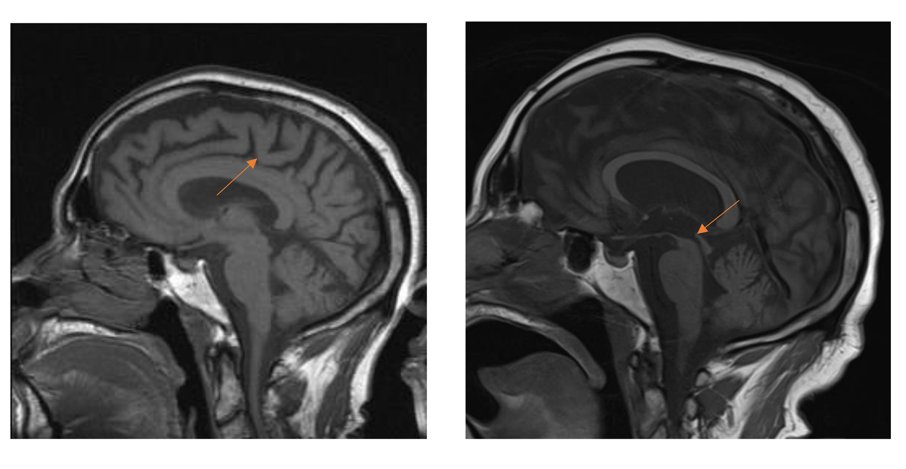
Figure 1. MRI T-1 weighted images, sagittal view. Left: Initial study eight years prior with greater midbrain and colliculus volume. Right: Current study demonstrating “hummingbird sign” (midbrain atrophy). Click to enlarge
Best-corrected visual acuities (BCVAs) were hand-motion OD and 20/50-1 OS. An afferent pupillary defect was noted OD. Evaluation of the patient’s extraocular muscle motility exhibited a complete restriction of inferior gaze and a moderate restriction of vertical gaze. Of particular note, the vestibulo-ocular reflex (VOR), or “doll’s head” reflex, was intact. The patient’s low phonation and mild dysphagia made it difficult to acquire a proper history to ascertain whether he had been symptomatic for his restriction of gaze.
Pre-dilated intraocular pressures (IOPs) were measured as 15 mmHg OD and 19 mmHg OS. Slit-lamp biomicroscopy revealed interpalpebral corneal punctate epithelial erosions OU, grade 2+ nuclear and anterior cortical cataracts OU with cortical spokes within the visual axis OD. Dilated fundus exam revealed cup-to-disk ratios of 0.6 OD and 0.4 OS with diffuse disc pallor OD and temporal disc pallor OS. The peripheral fundus exam was unremarkable OU. The patient had difficulty comprehending instructions for confrontational or kinetic visual fields.
The patient’s acquired oculomotor apraxia with intact VOR warranted further investigation to determine the presence or absence of a supranuclear palsy or degeneration. Magnetic resonance imaging (MRI) with and without contrast was ordered and revealed a classic “hummingbird sign” showing midbrain atrophy, as well as atrophy of the superior colliculus, which were not noted in the initial MRI study eight years prior (Figure 1). The patient’s primary care provider (PCP) was notified of the results, and a consultation to the neurology clinic for further evaluation of “possible PSP” was placed.
Case 2
A 61-year-old man presented for an eye examination reporting vertical binocular diplopia when reading for the past few weeks. The patient’s medical history included type 2 diabetes mellitus, hyperlipidemia, psoriasis and recently diagnosed PSP. Prior to the diagnosis of PSP, the patient had experienced frequent falls and generalized muscle weakness over the course of three years. Within the course of the three years, the patient had been prescribed carbidopa-levodopa (Sinemet) and his condition was treated as atypical Parkinson’s disease. At the time of his eye exam, he had discontinued carbidopa-levodopa due to a change in diagnosis from Parkinson’s disease to PSP. The patient’s ataxia had significantly progressed, and he was wheelchair-bound shortly before his eye examination. His most recent MRI demonstrated characteristic midbrain atrophy compared to prior studies (Figure 2). Current medications included acetaminophen 650 mg, aspirin 81 mg, atorvastatin 40mg, gabapentin 300 mg qam and 600 mg qhs, lorazepam 1 mg, metformin 500 mg, sertraline 150 mg and tizanidine 2 mg.

Figure 2. MRI, T-1 weighted images, sagittal view. Left: Initial study three years prior with robust midbrain. Right: Most recent study demonstrating significant midbrain atrophy. Click to enlarge
BCVAs were 20/20 OD and 20/20 OS. Pupils were equal, round and reactive to light with no afferent pupillary defect present. Confrontational fields were full to finger counting OD and OS. Pre-dilated IOPs were 16 mmHg and 14 mmHg. Slit-lamp biomicroscopy findings revealed unremarkable eyelid and eyelash findings, small nasal pterygium minimally encroaching on the cornea, flat and avascular iridis and deep and quiet angles OU. Dilated fundus exam revealed 1+ nuclear cataract OS, cup-to-disc ratios of 0.45 round and 0.5 round OD, OS, respectively, and otherwise unremarkable posterior pole and peripheral retinal findings. Based on evaluation of the patient’s vergences, exophoria greater at near than distance and a reduced near point of convergence were noted. Given the patient’s symptom of diplopia at near, he was diagnosed with convergence insufficiency secondary to PSP. He was prescribed 2.5 prism diopters base out in single-vision near glasses for greater comfort when reading.
Four months after the initial eye examination, the patient was admitted to a nursing home and was examined at bedside at the request of the PCP and charge nurse due to a reported increase of falls. The patient reported that he preferred to use previously prescribed progressive addition lenses despite having diplopia at near. Evaluation of the extraocular muscles revealed normal lateral gaze and intact VOR but limited superior and inferior movement. Due to these concerns, the patient was educated on the importance of protecting his eyes given his unstable gait and re-prescribed single-vision distance and single-vision near glasses with Fresnel prism to adjust for changes in severity of his diplopia.
Case 3
A 75-year-old male referred for an eye exam by his neurologist presented to the eye clinic for a preoperative cataract evaluation. Doctors were unable to elicit any verbal complaints from the patient, as he had severe aphasia rooting from his diagnosis of PSP. His wife stated he had been less interactive with the family, and she was concerned he was unable to see their faces as well as he used to. The patient’s neurologist had noted that improved vision may help reduce the possibility of agitation and delirium in demented patients. The patient’s medical history included chronic cough (from pooled airway secretions), chronic microaspirations and previous cerebral vascular accident with right arm contracture. His medications included levothyroxine 0.15 mg, memantine 5 mg, ferrous sulfate 300 mg/mL, omeprazole 40 mg, albuterol 05% 2.5 mg/0.5mL, bisacodyl 10 mg, ascorbic acid 250 mg, cholecalciferol 1,000 units, acetaminophen 650 mg, levetiracetam solution 125 mg/1.25mL and glycopyrrolate 1 mg.
BCVAs were unobtainable due to the patient’s poor cognitive state. Pupils were observed to be equal, round and reactive to light with no afferent pupillary defect present. The patient was unable to understand confrontational visual field testing. Upon extraocular muscle testing, he was noted to have restricted superior and inferior vertical gaze but intact VOR movement. Slit-lamp biomicroscopy findings revealed unremarkable eyelid and eyelash findings, flat and avascular iridis and deep and quiet angles OU. The lenses showed 3-4+ milky nuclear sclerotic cataracts OU. Dilated views by fundoscopy were poor and fleeting due to the patient’s tendency to squeeze his eyelids shut with light exposure. The retina was grossly normal with optic nerve cup-to-disc ratios possibly larger than average but difficult to ascertain.
The ophthalmologists gathered from the limited data that the patient would be able to cautiously proceed with cataract extraction pending presurgical medical evaluation. Evaluation by the patient’s pulmonologist determined that rhonchi, or low-pitched rattling sounds from the lungs, were present and suggested possible aspiration or respiratory infection. Given the patient’s demented mental status, general anesthesia would need to be administered, necessitating prolonged intubation and risking respiratory failure. Additionally, there was concern that coughing could disrupt postoperative sutures and result in a poor surgical outcome. The cataract surgery was thus postponed.
Education Guidelines
Learning objectives
- Understand neurodegenerative diseases as they relate to ocular health
- Become familiar with the ocular manifestations of PSP
- Understand possible complications of general anesthesia when considering ocular procedures in patients with PSP or other neurodegenerative disease affecting motor function
Key concepts
- Fundamental pathophysiology of PSP
- Ocular effects of PSP and the optometrist’s role in diagnosis
- Management of patients with PSP
Discussion points
- What is the pathophysiology of saccades and PSP?
- What causes PSP?
- How is PSP diagnosed?
- What are the ocular signs and symptoms of PSP?
- What are some differential diagnoses that should be considered in cases of suspected PSP?
- What is the appropriate management of patients with PSP?
- What is the prognosis of patients with PSP?
Literature review
PSP is the most common degenerative, atypical parkinsonian disorder, with a prevalence of 6.4 per 100,000 according to Schrag et al.5,13 The incidence is reported to increase with age from 1.7 cases per 100,000 at 50-59 years to 14.7 per 100,000 per year at 80-99 years.6,7 The mean age of diagnosis is approximately 65 years, with no racial or sex predilection. No significant risk factors for developing PSP have been identified.3
Discussion
Teaching instructions: Participants should read each question and consider how they would respond, then read the information provided in the text. Participants may work together in small groups or individually, either in real time or as part of a homework assignment. If working in groups, participants may split into three groups with each group focusing on one case or each group working on all three cases. Learning objectives can be assessed by comparing participants’ responses to the questions provided. This case may also be presented as a PowerPoint presentation detailing the case presentation, learning objectives, key concepts, literature review and discussion points.
What is the pathophysiology of saccades and PSP?
For horizontal and vertical saccades to be initiated, excitatory burst neurons (EBNs) in the brainstem generate a burst of neuronal discharge known as the pulse. The EBNs for horizontal saccades are located in the paramedian pontine reticular formation. The EBNs for vertical and torsional saccades are located in the rostral interstitial medial longitudinal fasciculus (RIMLF) and interstitial nucleus of Cajal. Once the pulse fires to the intended agonist muscle, antagonist muscles are relaxed.8
PSP results from an aggregation of abnormally phosphorylated tau proteins. Tau proteins aid in axonal transport and support neuronal microtubules. Localized accumulation of the irregular tau proteins form what are known as neurofibrillary tangles.9 In addition to the tauopathy, PSP degenerates dopaminergic neurons and cholinergic neurons, lending to loss of basal ganglia, cerebral cortex and, most clinically characteristic, brainstem structures. Structures within the brainstem that atrophy are the dorsal midbrain, notably the midbrain tegmentum and pedunculopontine nucleus, which lends to postural stability, and the motor nuclei of the cranial nerves in advanced stages of the disease. Given the atrophy of the midbrain tegmentum, the RIMLF is greatly affected, which decreases the presence of vertical EBNs, and an inability to initiate vertical saccades ensues.8
What causes PSP?
Although no definitive genetic factors have been identified, recent studies suggest there may be a genetic susceptibility in patients with mutations of the tau gene. PSP is currently considered a “tauopathy.”10
How is PSP diagnosed?
The diagnostic criteria for PSP, which were revised in 2017 by the International Parkinson and Movement Disorder Society, must include all “basic features”: sporadic occurrence, age 40 years or older at onset of first PSP-related symptom, and gradual progression of PSP-related symptoms.11,12 PSP-related symptoms include ocular motor dysfunction, postural instability, akinesia and cognitive dysfunction. The category of ocular motor dysfunction is further stratified to include vertical supranuclear gaze palsy, slow vertical saccades and frequent macro square wave jerks or “eyelid opening apraxia.” In this delineated criterion, the highest level of certainty for each category is defined as vertical supranuclear gaze palsy, repeated unprovoked falls within three years, progressive freezing of gait within three years, and speech or language disorder that presents as some variant of primary progressive aphasia, respectively. Supportive features that may increase diagnostic confidence, but do not alone suggest a diagnosis, include levodopa resistance, dysphagia, photophobia, and hypokinetic, spastic dysarthria.
Clinical forms of PSP have arisen since its original description in 1964. The original cases presented by Steele-Richardson-Olszewski have been classified as Richardson Syndrome (PSP-RS), which is characterized by postural instability, vertical gaze palsy and cognitive dysfunction. Eight other clinical variants have been described based on the severity and nature of their neurological signs.11
The gold standard for diagnosing PSP is a comprehensive, post-mortem neuropathological examination to identify the presence of neurofibrillary tangles. Because a definitive diagnosis requires a post-mortem exam, it is crucial for an eyecare provider to be able to identify oculomotor restrictions and make appropriate clinical recommendations for the patient’s systemic care.12
What are the ocular signs and symptoms of PSP?
Patients with PSP typically present with horizontal or vertical diplopia or asthenopia while reading secondary to convergence insufficiency, horizontal or vertical gaze palsy.13 Patients may also exhibit impaired slow phase responses of their vertical optokinetic response, as well as slowed volitional saccades.14 In a few studies, abnormal acoustic-startling reflex (orbicularis oculi response to high-intensity stimulation of the median nerve) and apraxia of eyelid opening have been observed in patients diagnosed with PSP.15-18 A decrease in blink frequency may cause symptoms of dry eye, including blurred vision, foreign body sensation, burning or irritation. Appropriate evaluation of extraocular muscle motility and near point of convergence as well as slit-lamp examination to evaluate the tear film and corneal integrity are of particular importance.
What are some differential diagnoses that should be considered in cases of suspected PSP?
From an ocular standpoint, it is important to determine the etiology of a patient’s presenting symptoms of double vision when reading or limitation of gaze rather than assuming such presentations are idiopathic or, as observed in case 2 reported above, presbyopia-associated convergence insufficiency. In assessing these complaints, the clinician must elicit a thorough case history including recent falls or postural changes and memory changes. Additionally, it is critical to carefully assess extraocular muscle movement, saccades, phorias and vergences at distance and near. Forced duction evaluation is particularly valuable in differentiating a mechanical vs. a neurodegenerative or vascular cause. The posterior thalamo-subthalamic paramedian artery, which stems from the posterior cerebral artery, supplies the RIMLF. Thus, an infarction of this artery may result in a superior gaze palsy.19 Neoplasms, particularly pineal gland tumors, may also lead to vertical gaze palsies.19 Other vertical gaze palsy conditions include Neimann-Pick type C, an autosomal-recessive condition in which cholesterol and lipids accumulate, dorsal midbrain syndrome, Whipple disease, and midbrain infarction.19
As previously noted, PSP is often misdiagnosed early in its course as Parkinson’s disease. It shows similar symptoms to Parkinson’s, dementia with Lewy bodies (DLB) and multiple system atrophy (MSA). Parkinson’s disease is a neurodegenerative disorder with characteristic skeletal muscle tremor, rigidity and akinesia, all of which are features observed in PSP.20 However, PSP typically involves greater cognitive dysfunction and speech disturbance than Parkinson’s disease. A study by Song et al. assessed patients with MSA, Parkinson’s disease and PSP. Results showed that 73% of patients with PSP had gaze abnormalities, a characteristic that was absent in Parkinson’s disease and MSA.21 Song also found that patients with PSP and MSA had a poorer response to levodopa, which is a staple treatment for Parkinson’s disease.
MSA is defined as an “adult-onset, sporadic, progressive neurodegenerative disease” with “parkinsonian features, cerebellar ataxia, autonomic failure, urogenital dysfunction, and corticospinal disorders” by the Second Consensus Statement on the Diagnosis of MSA.22,23 Although difficult to definitively differentiate from PSP, any sign of autonomic dysfunction would steer the diagnosis towards MSA.
DLB is defined by the 2017 Dementia with Lewy Bodies Consortium as a disease of “progressive cognitive decline of sufficient magnitude to interfere with normal social or occupational functions, or with usual daily activities.” In DLB, patients have “recurrent visual hallucinations that are typically well-formed and detailed,” rapid eye movement (REM) sleep behavior disorder, and “features of parkinsonianism.”24 Gait abnormalities, gaze apraxia and saccadic dysfunction may be present in both PSP and DLB, but visual hallucinations are a hallmark feature of DLB.25,26
Various presenting signs and symptoms overlap between PSP and other neurodegenerative disorders, which makes PSP diagnosis reliant on the clinical findings centered around the 2017 Movement Disorder Society criteria. Neuroimaging such as MRI may be a helpful supplement for diagnosis. Various studies report abnormalities on MRI demonstrating midbrain atrophy, third ventricle dilation, T2-periaqueductal hyperintensities and frontal and temporal atrophy. One hallmark feature of PSP on MRI is the “hummingbird sign” or “penguin sign” demonstrating midbrain atrophy. Most useful for differentiating PSP from Parkinson’s disease or other atypical Parkinson’s diseases are the midbrain-to-pontine area ratio, which tends to be lower in PSP patients, and the magnetic resonance parkinsonism index (MRPI), which is greater in PSP patients. The MRPI is a value calculated by determining atrophy of the midbrain, superior cerebellar peduncle, pons and middle cerebellar peduncle.27,28 Another promising diagnostic method is utilizing positron emission tomography scanning to track the tau protein THK5351 and determine the presence of tau aggregates specific to PSP.29 This diagnostic tool, however, is predominantly used in research and often not clinically performed due to cost constraints.
What is the appropriate management of patients with PSP?
Eyecare providers are crucial in determining the presence or absence of gaze palsies, which aids in the differentiation of PSP. Once an abnormality is determined, a referral to the patient’s PCP and neurologist should be made.
Patients with PSP often have limited benefit from levodopa therapy, as opposed to patients with Parkinson’s disease. In a study by Williams et al. investigating 91 pathologically confirmed cases of PSP, 32% of patients presented with a response to levodopa, defined as a 30% or greater improvement in symptoms.30 There are no successful pharmacological treatment options targeting the disease process, and management centers around symptomatic care. A few case reports have demonstrated some efficacy of botulinum toxin injected into the orbicularis oculi for apraxia of eyelid opening, into upper limbs to improve rigidity and into cricopharyngeal muscle for dysphagia.31-34 Additional intervention from an interdisciplinary care team consisting of speech, physical and occupational therapists is necessary to facilitate greater independent activities of daily living.
Following diagnosis of PSP, the optometrist may play a role in the patient’s care, particularly to address diplopia and dry eye symptoms. A study by Reddy et al. found that corneal sensitivity was reduced in patients with PSP, with 71% reporting they did not have dry eyes. However, evaluation of the tear firm revealed reduced tear break-up time compared to the control group.35 Even without patent report of dry eye symptoms, it is important for the optometrist to manage the patient’s dry eyes with artificial tears and lubricating ointment or gel formulations for severe exposure keratopathy. Although few reports demonstrate alleviation of double vision when reading with “mirror prism,” we found that Fresnel prisms were particularly useful for alleviating the progressive double vision experienced by the patient in case 2.36,37
What is the prognosis for patients with PSP?
PSP is a rapidly neurodegenerative condition with a poor prognosis. A study by Cosseddu et al. evaluated 100 patients with PSP and found that the average disease duration following diagnosis was 8.25 years.3 Cosseddu found that patients with dementia at the time of diagnosis had a shorter survival time than those without dementia, with no other significant predictors.3 The most common causes of death for patients with PSP are respiratory-related, with the most frequent complication being aspiration pneumonia.38 Given the high frequency of respiration complications, surgery requiring general anesthesia should be given significant consideration due to the potential for respiratory failure.
Conclusion
Optometrists may play a critical role in recognizing characteristic features of PSP and managing concomitant ocular symptoms that may arise. Although no pharmacologic therapy halts progression of the disease, a multidisciplinary team can provide patients and their caretakers with the tools to maximize quality of life and minimize debilitating symptoms.
References
- Steele JC, Richardson JC, Olszewski J. Progressive supranuclear palsy. A heterogeneous degeneration involving the brain stem, basal ganglia and cerebellum with vertical gaze and pseudobulbar palsy, nuchal dystonia and dementia. Arch Neurol. 1964 Apr;10:333-59.
- Joutsa J, Gardberg M, Röyttä M, Kaasinen V. Diagnostic accuracy of Parkinsonism syndromes by general neurologists. Parkinsonism Relat Disord. 2014 Aug;20(8):840-4.
- Cosseddu M, Benussi A, Gazzina S, et al. Natural history and predictors of survival in progressive supranuclear palsy. J Neurol Sci. 2017 Nov 15;382:105-107.
- Glasmacher SA, Leigh PN, Saha RA. Predictors of survival in progressive supranuclear palsy and multiple system atrophy: a systematic review and meta-analysis. J Neurol Neurosurg Psychiatry. 2017 May;88(5):402-411.
- Schrag A, Ben-Shlomo Y, Quinn NP. Prevalence of progressive supranuclear palsy and multiple system atrophy: a cross-sectional study. Lancet. 1999 Nov 20;354(9192):1771-5.
- Bower JH, Maraganore DM, McDonnell SK, Rocca WA. Incidence of progressive supranuclear palsy and multiple system atrophy in Olmsted County, Minnesota, 1976 to 1990. Neurology. 1997 Nov;49(5):1284-8.
- Chiu WZ, Kaat LD, Seelaar H, et al. Survival in progressive supranuclear palsy and frontotemporal dementia. J Neurol Neurosurg Psychiatry. 2010 Apr;81(4):441-5.
- Kato N, Arai K, Hattori T. Study of the rostral midbrain atrophy in progressive supranuclear palsy. J Neurol Sci. 2003;210(1-2):57-60.
- Bancher C, Lassmann H, Budka H, et al. Neurofibrillary tangles in Alzheimer’s disease and progressive supranuclear palsy: antigenic similarities and differences. Microtubule-associated protein tau antigenicity is prominent in all types of tangles. Acta Neuropathol. 1987;74(1):39-46.
- Stanford PM, Halliday GM, Brooks WS, et al. Progressive supranuclear palsy pathology caused by a novel silent mutation in exon 10 of the tau gene: expansion of the disease phenotype caused by the tau gene mutations. Brain. 2000 May;123 (Pt 5):880-93.
- Höglinger GU, Respondek G, Stamelou M, et al. Clinical diagnosis of progressive supranuclear palsy: the movement disorder society criteria. Mov Disord. 2017 Jun;32(6):853-864.
- Litvan I, Agid Y, Calne D, et al. Clinical research criteria for the diagnosis of progressive supranuclear palsy (Steele-Richardson-Olszewski syndrome): report of the NINDS-SPSP international workshop. Neurology. 1996 Jul;47(1):1-9.
- Williams DR, Lees AJ. Progressive supranuclear palsy: clinicopathological concepts and diagnostic challenges. Lancet Neurol. 2009 Mar;8(3):270-9.
- Chen AL, Riley DE, King SA, et al. The disturbance of gaze in progressive supranuclear palsy: implications for pathogenesis. Front Neurol. 2010 Dec 3;1:147.
- Shaikh AG, Factor SA, Juncos J. Saccades in progressive supranuclear palsy–maladapted, irregular, curved, and slow. Mov Disord Clin Pract. Sep-Oct 2017;4(5):671-681.
- Gironell A, Kulisevsky J, Roig C, Pascual-Sedano B, Rodríguez-Fornells A, Otermin P. Diagnostic potential of acoustic startle reflex, acoustic blink reflex, and electro‐oculography in progressive supranuclear palsy: a prospective study. Mov Disord. 2003 Nov;18(11):1273-9.
- Valls-Solé J, Valldeoriola F, Tolosa E, Marti MJ. Distinctive abnormalities of facial reflexes in patients with progressive supranuclear palsy. Brain. 1997 Oct;120 (Pt 10):1877-83.
- Grandas F, Esteban A. Eyelid motor abnormalities in progressive supranuclear palsy. In: Tolosa E, Duvoisin R, Cruz-Sanchez FF, editors. Progressive supranuclear palsy: diagnosis, pathology, and therapy. Vienna: Springer;1994. p.33-41.
- Lloyd-Smith Sequeira A, Rizzo JR, Rucker JC. Clinical approach to supranuclear brainstem saccadic gaze palsies. Front Neurol. 2017 Aug 23;8:429.
- Lopez G, Bayulkem K, Hallett M. Progressive supranuclear palsy (PSP): Richardson syndrome and other PSP variants. Acta Neurol Scand. 2016 Oct;134(4):242-9.
- Pasternak JJ, Lanier WL. Diseases affecting the brain. In: Hines RL, Marschall KE, editors. Stoelting’s anesthesia and co-existing disease. Philadelphia: Elsevier; 2018. p. 265-303.
- Williams DR, Warren JD, Lees AJ. Using the presence of visual hallucinations to differentiate Parkinson’s disease from atypical parkinsonism. J Neurol Neurosurg Psychiatry. 2008 Jun;79(6):652-5.
- Gilman S, Wenning GK, Low PA, et al. Second consensus statement on the diagnosis of multiple system atrophy. Neurology. 2008 Aug 26;71(9):670-6.
- Liscic RM, Srulijes K, Gröger A, Maetzler W, Berg D. Differentiation of progressive supranuclear palsy: clinical, imaging and laboratory tools. Acta Neurol Scand. 2013 May;127(5):362-70.
- Song YJ, Huang Y, Halliday GM. Clinical correlates of similar pathologies in parkinsonian syndromes. Mov Disord. 2011 Feb 15;26(3):499-506.
- McKeith IG, Boeve BF, Dickson DW, et al. Diagnosis and management of dementia with Lewy bodies: fourth consensus report of the DLB Consortium. Neurology. 2017 Jul 4;89(1):88-100.
- Hussl A, Mahlknecht P, Scherfler C, et al. Diagnostic accuracy of the magnetic resonance parkinsonism index and the midbrain‐to‐pontine area ratio to differentiate progressive supranuclear palsy from Parkinson’s disease and the Parkinson variant of multiple system atrophy. Mov Disord. 2010 Oct 30;25(14):2444-9.
- Massey LA, Jager HR, Paviour DC, et al. The midbrain to pons ratio: a simple and specific MRI sign of progressive supranuclear palsy. Neurology. 2013 May 14;80(20):1856-61.
- Respondek G, Levin J, Höglinger GU. Progressive supranuclear palsy and multiple system atrophy: clinicopathological concepts and therapeutic challenges. Curr Opin Neurol. 2018 Aug;31(4):448-454.
- Williams DR, Silva R, Paviour DC, et al. Characteristics of two distinct clinical phenotypes in pathologically proven progressive supranuclear palsy: Richardson’s syndrome and PSP-parkinsonism, Brain. 2005 Jun;128 (Pt 6):1247-58.
- Lepore V, Defazio G, Acquistapace D, et al. Botulinum a toxin for the so‐called apraxia of lid opening. Mov Disord. 1995 Jul;10(4):525-6.
- Piccione F, Mancini E, Tonin P, Bizzarini M. Botulinum toxin treatment of apraxia of eyelid opening in progressive supranuclear palsy: report of two cases. Arch Phys Med Rehabil. 1997 May;78(5):525-9.
- Polo KB, Jabbari B. Botulinum toxin‐A improves the rigidity of progressive supranuclear palsy. Ann Neurol. 1994 Feb;35(2):237-9.
- Alfonsi E, Merlo IM, Ponzio M, et al. An electrophysiological approach to the diagnosis of neurogenic dysphagia: implications for botulinum toxin treatment. J Neurol Neurosurg Psychiatry. 2010 Jan;81(1):54-60.
- Reddy VC, Patel SV, Hodge DO, Leavitt JA. Corneal sensitivity, blink rate, and corneal nerve density in progressive supranuclear palsy and Parkinson disease. Cornea. 2013 May;32(5):631-5.
- Krohel GB, Tobin DR, Hartnett ME, Barrows NA. Divergence paralysis. Am J Ophthalmol. 1982 Oct;94(4):506-10.
- Weisz RR, Richer SP, Jordan M, Tucker EG. Case report: prismatic redirection of vision for progressive supranuclear palsy. J Am Optom Assoc. 1991 Jul;62(7):537-9.
- Litvan I, Mangone CA, McKee A, et al. Natural history of progressive supranuclear palsy (Steele-Richardson-Olszewski syndrome) and clinical predictors of survival: a clinicopathological study. J Neurol Neurosurg Psychiatry. 1996 Jun;60(6):615-20.

