Multiple Evanescent White Dot Syndrome in an Atypical Patient
Melissa Tawa, OD, FAAO
Multiple evanescent white dot syndrome (MEWDS) is a rare, self-limiting inflammation of the retinal pigment epithelium and outer photoreceptors. It is characterized by multiple white dots in the posterior pole and mid-periphery and foveal granularity. Most patients have spontaneous resolution within weeks. This teaching case report reviews the epidemiology, pathophysiology, differential diagnoses and management of MEWDS. Instructors are guided on how to use the case in educational settings, such as small group discussions in the classroom or clinic. Although MEWDS is a relatively rare condition, this teaching case report challenges learners clinical reasoning skills and multimodal imaging interpretation.
Keywords
Introduction
Multiple evanescent white dot syndrome (MEWDS) is a rare white dot syndrome characterized by white dots in the posterior pole and mid-periphery, and foveal granularity.1 It was previously thought to be a disease of the choriocapillaris, however more recent multimodal imaging studies suggest that the retinal pigment epithelium-photoreceptor complex may be the primary site of inflammation.2 It typically affects young adult myopic females between 20 to 40 years of age3 and may be preceded by a flu-like viral episode.4 Patients classically present with complaints of decreased visual acuity and photopsias.3 MEWDS is typically a self-limiting condition.2
This teaching case report aims to facilitate discussion around the clinical presentation of MEWDS. While MEWDS is a relatively rare condition and the atypical patient demographics makes this case more challenging; it is an exercise in clinical reasoning skills and an opportunity to discuss various multimodal imaging techniques frequently used in more common posterior segment diseases.
Case Description
Visit 1
A 16-year-old Latino male presented for a routine comprehensive eye examination. He denied any known medical conditions, a denial confirmed by the patient’s father. His ocular history was significant for longstanding chorioretinal scars in the left eye. He reported no ocular complaints with his current glasses. Best corrected visual acuity was 20/20 in each eye with mixed astigmatism in the right eye and compound hyperopic astigmatism in the left. Pupils, extraocular muscle movements and confrontation visual fields were within normal limits in both eyes. Anterior segment findings were normal in each eye. Intraocular pressures were 18 mmHg in both the right and left eye. He declined dilation that day and scheduled a dilated fundus examination in 2 weeks. Posterior segment examination was normal to the extent seen undilated. Widefield color photography (Figures 1a and 1b) was also normal to the extent imaged in each eye.
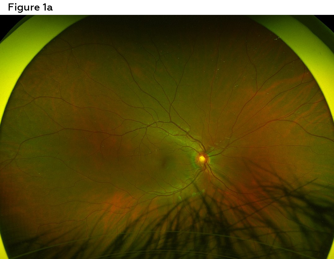
Figure 1a. Widefield color fundus photo right eye, visit one. No retinopathy or optic neuropathy. Click to enlarge
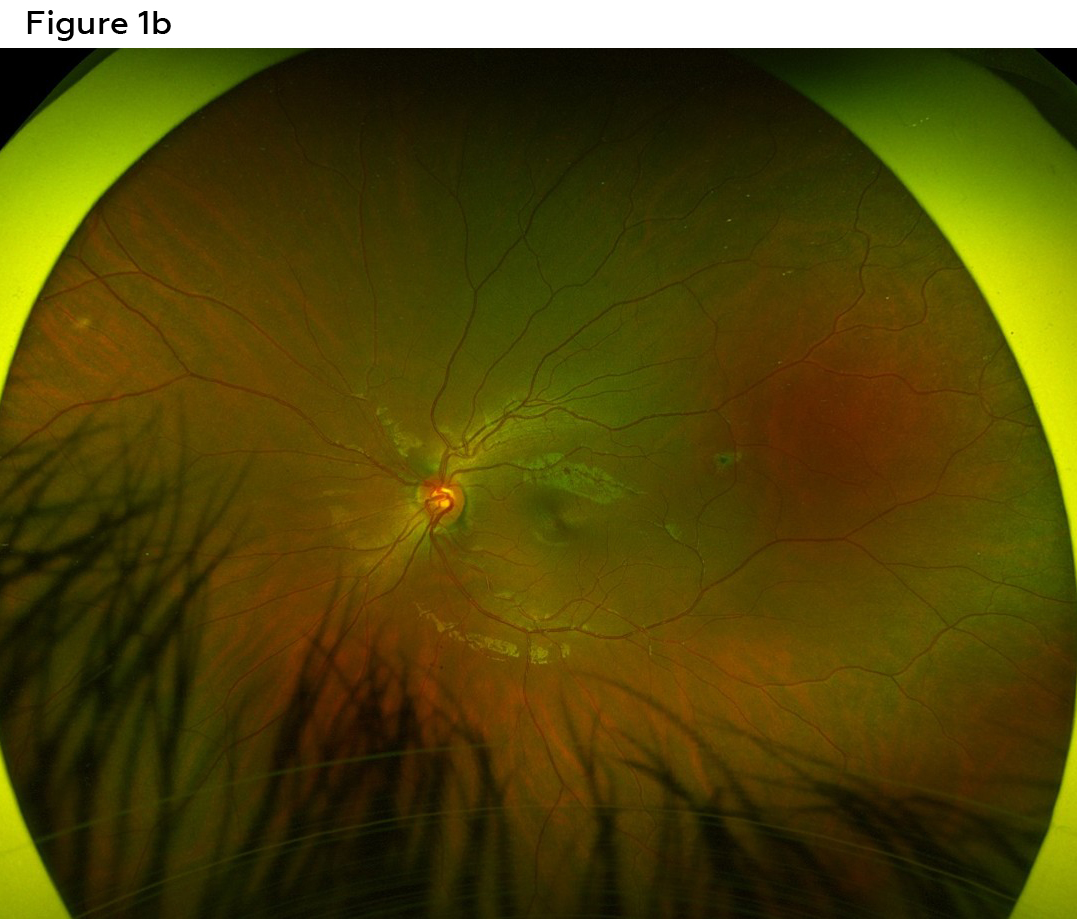
Figure 1b. Widefield color fundus photo left eye, visit one. Aside from the longstanding chorioretinal scar at the end of the superotemporal arcades, there is no other retinopathy or optic neuropathy. Click to enlarge
Visit 2
The patient returned 2 weeks later for his scheduled dilation. He reported that the day after his comprehensive examination, he visited a theme park and rode multiple rollercoasters. He denied any direct trauma to his head or eyes but noted that he experienced significant head movement on the rollercoasters. That afternoon, while at the theme park, he reported noticing flashes and floaters in his right eye. He also reported that the quality of vision in the right eye was not as good as the left, particularly in dim lighting.
Best corrected visual acuity remained 20/20 in each eye. Pupils, extraocular muscle movements and confrontation visual fields were within normal limits in each eye. Anterior segment findings were normal in each eye. Intraocular pressures were 20 and 19 mmHg right eye and left eye, respectively. The patient was dilated with tropicamide 1% and phenylephrine 2.5% in both eyes. Given the history of rollercoasters and symptoms of flashes and floaters, careful attention was paid to signs of retinal tears/detachments. Evaluating the anterior vitreous revealed no Shafer sign present in either eye. Posterior segment evaluation revealed many small hypopigmented lesions around the vascular arcades in the posterior pole, more prominent superiorly and extending towards the equator of the right eye. Careful examination of the fovea with slit lamp fundus biomicroscopy evaluation revealed subtle granularity in the right eye only. The optic nerves had distinct margins and healthy rim tissue in both eyes. There were no holes, breaks, tears or detachments in either eye. The posterior segment findings of the left eye were unremarkable. Widefield color photos from this visit can be seen in Figures 2a and 2b.
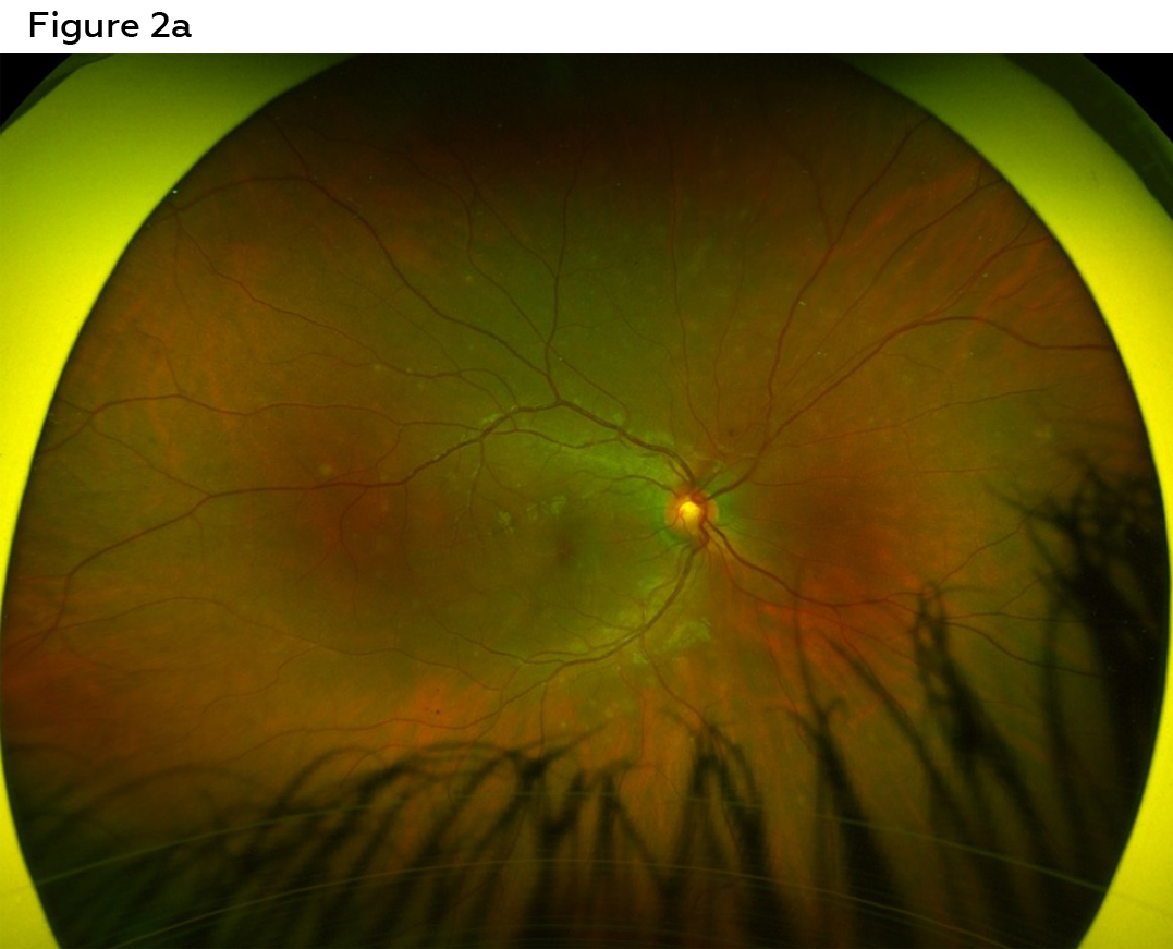
Figure 2a. Widefield color fundus photo right eye, visit two. White round lesions can be seen in the posterior pole and equator, greatest in the superior hemifield. Click to enlarge
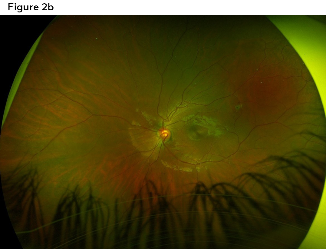
Figure 2b. Widefield color fundus photo left eye, visit two. Left eye is stable when compared to the visit 2 weeks prior without the presence of white round lesions or other retinopathy. Click to enlarge
Visit 3
Due to insurance, the patient’s father requested a referral to an in-network provider for the follow-up visit. The patient was seen by an outside provider 6 weeks later with full resolution of both symptoms and posterior segment findings in the right eye. There was no new retinopathy or other significant ocular findings in either eye.
Preliminary Differential Diagnoses
Trainees should be asked to pause and come up with preliminary differential diagnoses based on the information gathered so far, as these will guide further testing in the next section. Corrective feedback should not be given at this time since trainees will have the opportunity to re-evaluate differential diagnoses after the results of multimodal imaging are presented. Examples of reasonable differential diagnoses include the following:
- Retinal tear/detachment: Based on the patient’s chief complaint of flashes and floaters and the history of riding rollercoasters at the onset of symptoms, a differential diagnosis of a retinal tear or detachment was considered early in the examination. However, careful dilated evaluation revealed no tears or detachments in the retina and no Shafer sign in the anterior vitreous. This differential diagnosis was ruled out prior to imaging.
- Commotio retinae
- Multiple evanescent white dot syndrome
- Other white dot syndromes – ex: acute posterior multifocal placoid pigment epitheliopathy (APMPPE), idiopathic multifocal choroiditis, acute zonal occult outer retinopathy (AZOOR), birdshot chorioretinopathy, serpiginous chorioretinopathy
- Masqueraders of white dot syndromes – ex: syphilitic outer retinitis, primary vitreoretinal lymphoma
Additional Testing
Based on the patient’s presentation, trainees can now be asked about additional tests they would perform to aid in the diagnosis of the patient. Emphasis should be placed on testing that will be most discriminatory in the diagnosis of this patient’s condition. Trainees should also be specific in the types of tests ordered (ex: macula optical coherence tomography instead of optical coherence tomography, or threshold visual field instead of visual field). Residents or more advanced students can take it one step further and be asked about the expected imaging results for their differential diagnoses. Then, once the trainee has requested testing, they can then be shown the results provided in this teaching case report and be asked to interpret the data.
Fundus autofluorescence (Figures 3a and 3b) was performed to further evaluate the white lesions seen on color fundus photography and clinical exam. Fundus autofluorescence revealed patchy hyperautofluorescence in the peripapillary region, greatest superiorly, as well as paramacular region and into the arcades corresponding with the hypopigmented lesions seen clinically in the right eye. The areas of hyperautofluorescence exceeded the number of white dots seen clinically, particularly in the peripapillary region. Fundus autofluorescence findings were normal for the left eye.
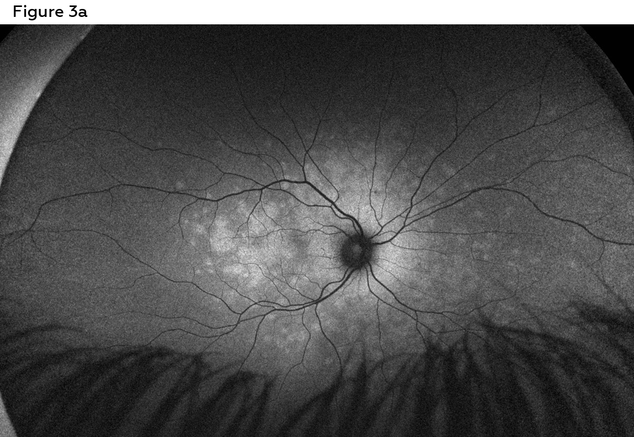
Figure 3a. Widefield color fundus photo right eye, visit two. Patchy hyperautofluorescence in the peripapillary region, greatest superiorly. Hyperautofluorescence is also seen in the paramacular region. Click to enlarge
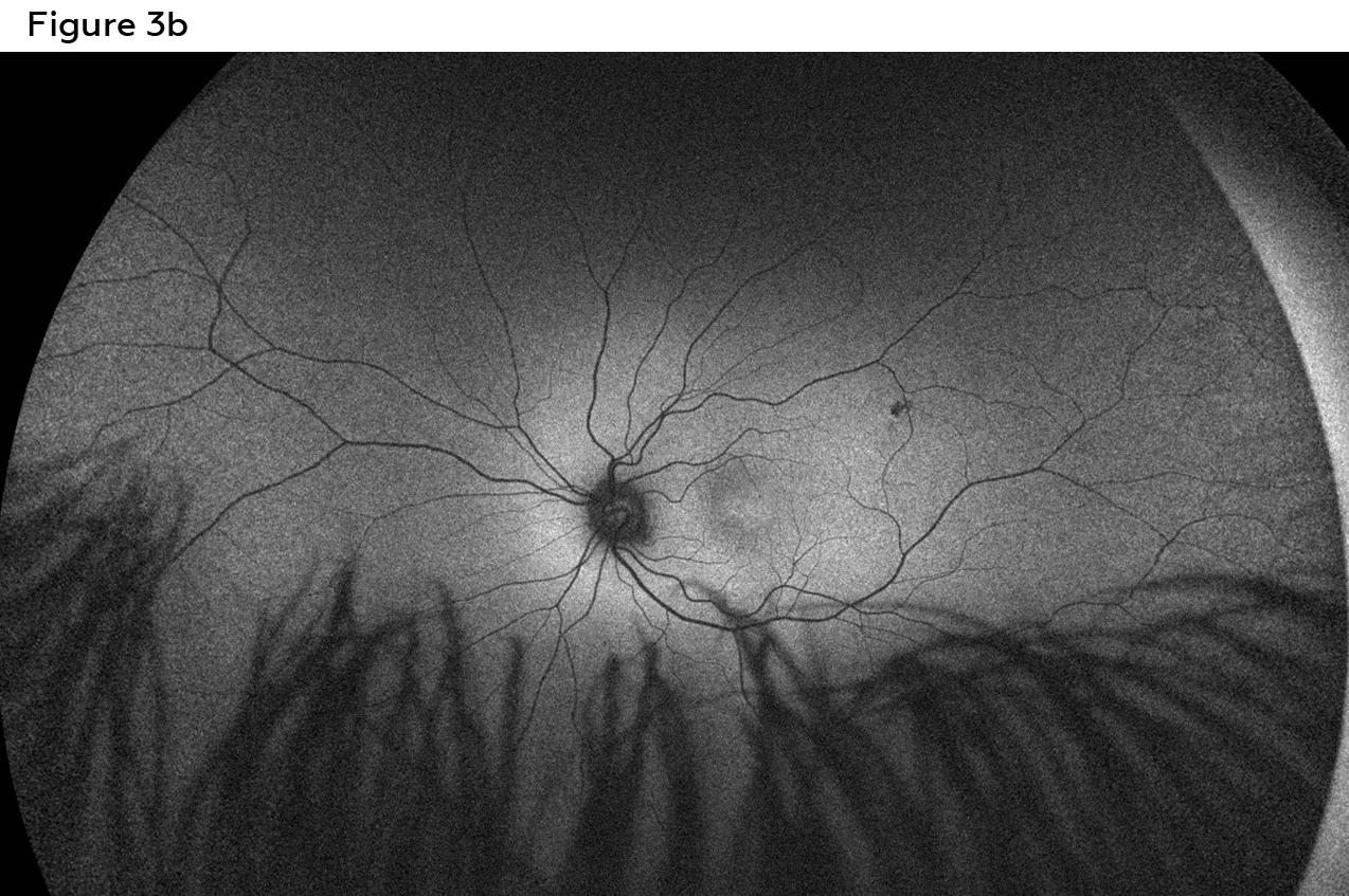
Figure 3b. Widefield color fundus photo left eye, visit two. Normal fundus autofluorescence pattern aside from the hypoautofluorescence of the longstanding chorioretinal scar. Click to enlarge
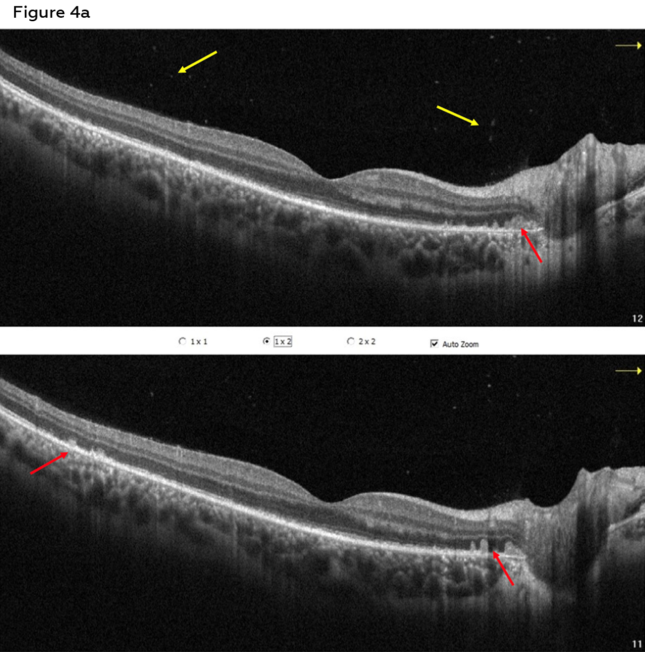
Figure 4a. Macula OCT right eye, visit two. Raster of the right eye shows loss of the ellipsoid zone just temporal to the fovea and retinal pigment epithelium inflammation (red arrows). Mild vitreous cells are present (yellow arrows). Click to enlarge
Macula optical coherence tomography (OCT) (Figures 4a and 4b) was performed for both eyes to look for correlations with the white dots and fundus autofluorescence findings as well as the foveal granularity and complaint of reduced quality of vision in the right eye. Right eye OCT revealed mild vitritis (yellow arrows). There were also several areas of retinal pigment epithelium inflammation (red arrows), as represented by the hyperreflective lesions of the retinal pigment epithelium extending anteriorly into the outer nuclear layer. These lesions are particularly evident in the temporal peripapillary region and correspond to the hypopigmented lesions seen on clinical examination and hyperautofluorescence on fundus autofluorescence. Disruption of the foveal ellipsoid zone can also be seen in the right temporal subfoveally and into the temporal parafoveal region. Macula OCT findings were normal for the left eye.
Macula OCT of the right eye without arrows can be found in the Appendix and should be used to show trainees when they request additional testing. This version with arrows can be used to point out the vitritis and RPE inflammation if missed during the initial discussion.
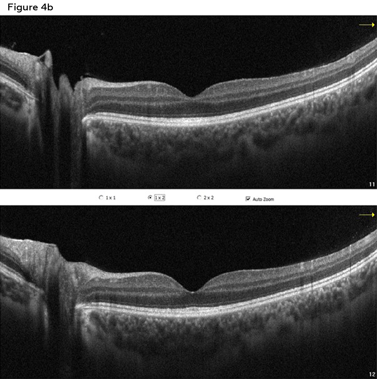
Figure 4b. Macula OCT left eye, visit two. Normal macula OCT findings of the left eye. The ellipsoid zone is well defined and fully intact. Click to enlarge
A Humphrey Visual Field 24-2 SITA Faster (Figures 5a and 5b) was performed on both eyes. Results showed a slightly reduced foveal threshold in the right eye (34 dB) with an enlarged blind spot and moderate paracentral defects greater superiorly than inferiorly. These defects correspond with the hyperautofluorescent areas seen in fundus autofluorescence. They also account for his complaint of decreased quality of vision in the right eye. The visual field 24-2 for the left eye had a few mild non-contiguous defects, unremarkable for neurologic or ocular disease. It is appropriate for trainees to request a Humphrey Visual Field 30-2, full-field or other similar threshold testing strategy. A Humphrey Visual Field 10-2 or microperimetry may be done to look at central fixation function; however, they should be performed in conjunction with wider field threshold testing strategies, as the fundus autofluorescence and white dots on clinical exam indicate involvement outside of the central ten degrees.
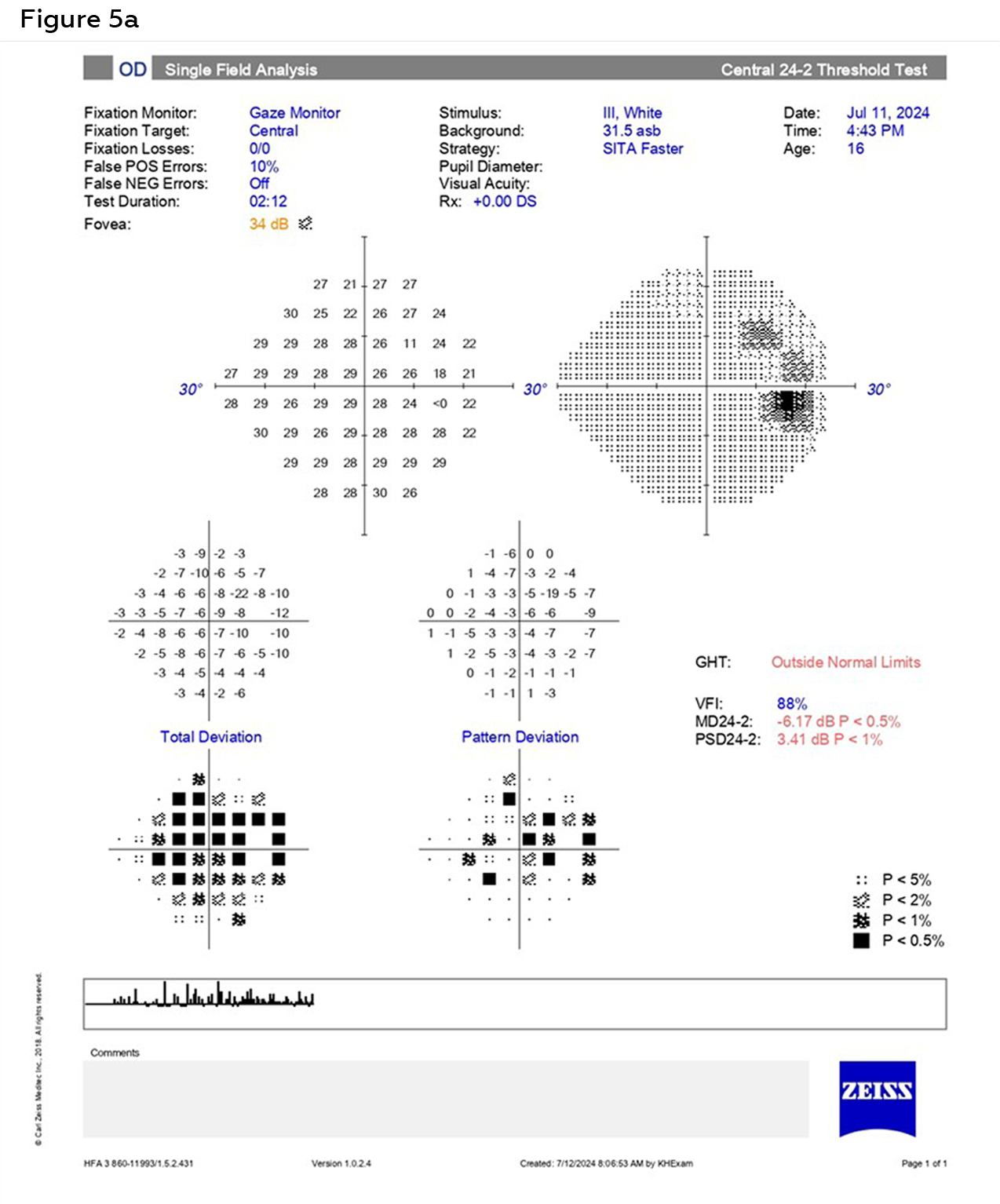
Figure 5a. Humphrey Visual Field 24-2 right eye, visit two. Visual field findings indicate a slightly reduced foveal threshold, enlarged blind spot and moderate paracentral defects, greater superiorly than inferiorly. Click to enlarge
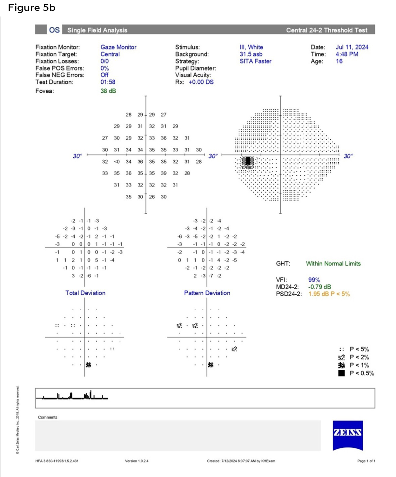
Figure 5b. Humphrey Visual Field 24-2 left eye, visit two. The visual field defects are mild and mostly edge points. There is no suggestion of neurologic, glaucomatous or retinal pathology. Click to enlarge
Updated Differential Diagnoses
Now that imaging has been obtained, trainees should be asked to come up with their updated differential diagnoses. Trainees should also be asked to explain case findings that are consistent and/or inconsistent with each of the proposed differentials to demonstrate clinical reasoning skills. This exercise should be a formative assessment, where knowledge gaps are identified, and feedback provided throughout the discussion. Table 1 compares the three white dot syndromes listed in the differential diagnoses and can be used to facilitate discussion about this group of conditions.
- Multiple evanescent white dot syndrome: MEWDs belongs to the white dot syndromes and typically affects young adult myopic females between 20 to 40 years of age.1,3 The disease is characterized by white dots in the posterior pole and mid-periphery, mild vitritis and foveal granularity.1 Patients classically present with complaints of decreased visual acuity and photopsias.3 The retinal pigment epithelium-photoreceptor complex may be the primary site of inflammation. Ocular signs and symptoms may be preceded by a flu-like viral episode.4 MEWDS is typically a self-limiting condition.2
- Commotio retinae: Commotio retinae is described as retinal whitening after blunt trauma to the globe. This deep retinal whitening occurs opposite the site of impact. Macular edema, also known as Berlin’s edema, may mimic a cherry red spot, which is classically associated with a central retinal artery occlusion.5 Expected OCT findings include increased reflectivity of the ellipsoid zone, disruption of the interdigitation zone and loss of reflectivity of the external limiting membrane.6 Vision and edema typically resolve over several weeks and can leave behind granular hyperpigmentation.5
- Acute posterior multifocal placoid pigment epitheliopathy (APMPPE): APMPPE is another white dot syndrome. It occurs in adults, often between 20 to 50 years of age, is typically bilateral, and patients often complain of reduced vision. Flat, round and confluent cream-colored plaque-like lesions with indistinct margins can be seen in the posterior pole, representing subretinal infiltrates. Indocyanine green angiography and OCT angiography have demonstrated that inner choroidal ischemia is the underlying cause of disease. These lesions resolve on their own and may leave behind hypopigmentation and later pigment mottling or clumping. If the placoid lesions and choroidal ischemia persist for more than 3 months, the condition is referred to as persistent placoid maculopathy.5
- Idiopathic multifocal choroiditis: Idiopathic multifocal choroiditis also belongs to the white dot syndromes and is an inflammatory choroiditis. It tends to affect myopic women between 20 to 60 years with reported symptoms of photopsias and blurry vision (more than half have visual acuity worse than 20/100 in the worse seeing eye).5, 7 In the acute phase, round or oval yellow lesions are seen in the posterior pole, peripapillary region and midperiphery. They may form a linear pattern and be associated with subretinal fluid. It is commonly bilateral and associated with anterior chamber and vitreous cells ranging from quiescent to severe. The yellow lesions can later develop into atrophic chorioretinal scars with pigmentation, and choroidal neovascular membranes commonly develop. The clinical course tends to wax and wane but can be chronic in some cases.5
- Syphilitic outer retinitis: Syphilis is known as the “great masquerader” of ocular diseases, including MEWDS. Syphilitic outer retinitis can present subtly, but characteristic features include flat yellow lesions of the outer retina, disruption of the ellipsoid zone and increased fundus autofluorescence (FAF) signal. Phlebitis may also be a key feature of syphilitic outer retinitis (SOR) and is well observed with fluorescein angiography.8
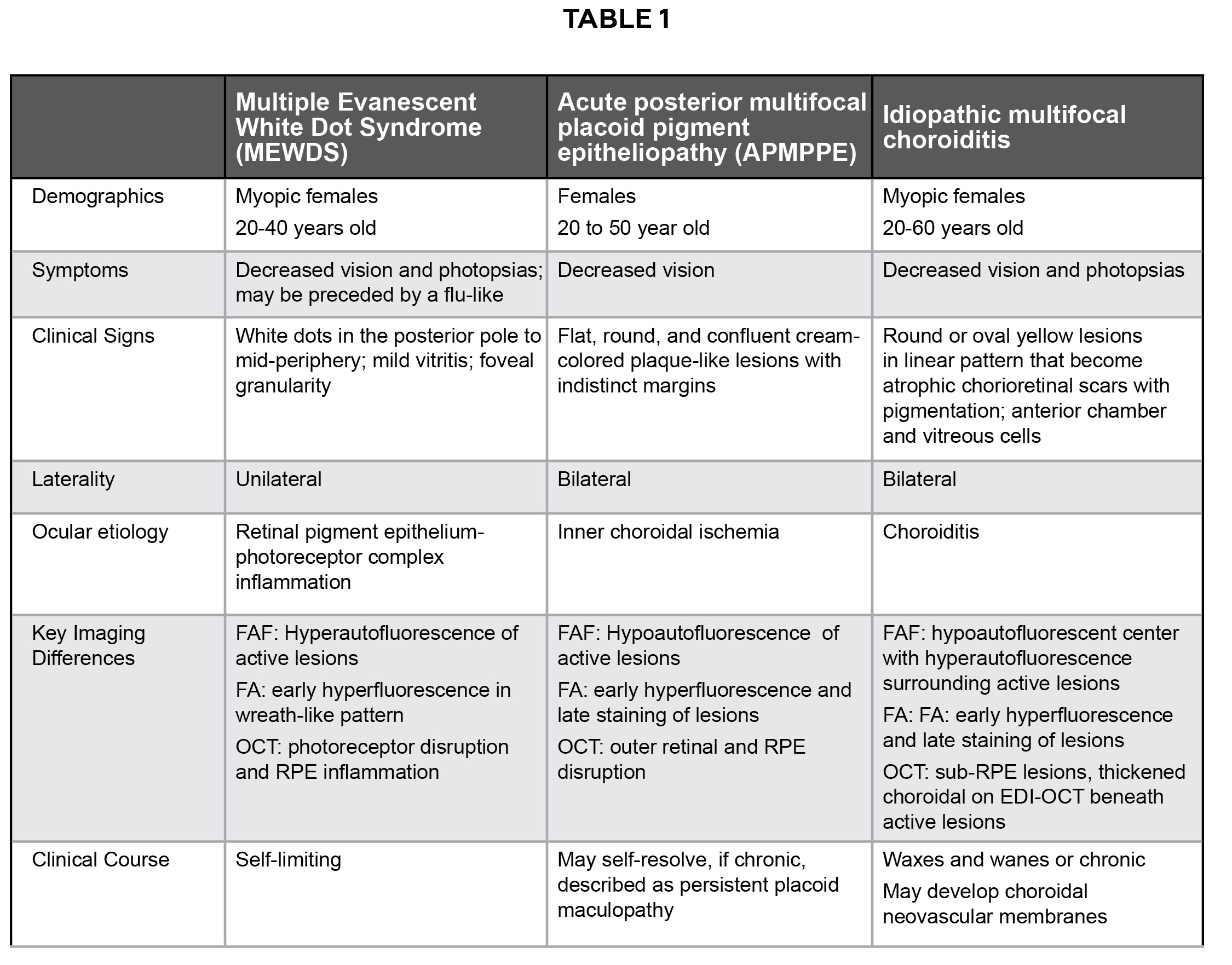
Table 1. Summary table of common findings of the three white dot syndromes in the differential diagnoses. Click to enlarge
Additional History
After interpretation of multimodal imaging on visit two, the patient was asked further case history questions to narrow down the differential diagnoses. Because MEWDS may be preceded by a flu-like viral episode,4 the patient was asked about flu-like symptoms. He reported that he was training for wrestling and is often fatigued. He reported that it was possible he would not recognize mild flu-like symptoms, thinking they are part of his training fatigue. He also denied a recent diagnosis of COVID-19 infection as well as any other vaccinations. The patient reported a normal routine physical examination within the school year. He also denied any new systemic signs or symptoms aside from his usual wrestling fatigue, including headaches, skin rashes or skin lesions. The patient reported that he has never been sexually active.
Final Diagnosis
During or after the differential diagnoses are discussed, the trainee should be asked to come up with a final diagnosis based on the findings of the case.
Given the history of roller coaster rides coinciding with the onset of symptoms, ocular trauma, such as retinal tear/detachment and commotio retinae were differential diagnoses. Earlier trainees may focus on this aspect of the case and limit their differential diagnoses to trauma-related conditions. However, upon further questioning the patient denied any direct contact to his head or eyes while riding the rollercoasters and dilated ocular evaluation revealed no retinal tears, breaks or detachments. The lack of direct trauma also helps rule out commotio retinae. Because the white dots were in the posterior pole, and in commotio retinae, the retinal whitening is opposite the site of impact, he would have had to experience trauma to the anterior portion of his eye/globe. Additionally, he lacked the classic confluent retinal whitening and edema associated with commotio retinae. Instead of the expected increased reflectivity of the ellipsoid zone with commotio retinae, the patient in this case displayed disruption of the ellipsoid zone, and there was no decrease in external limiting membrane reflectivity compared to the normal left eye on macula OCT.
While the typical patient diagnosed with multiple evanescent white dot syndrome is a young adult myopic female, this patient was slightly younger as a teenage male with minimal myopia in the right eye only. His racial background is not pertinent to this case as MEWDS lacks known racial associations. However, his ocular examination and multimodal imaging findings aligned best with a diagnosis of MEWDS in the right eye. Key distinguishing features include unilateral presentation, white dots, foveal granularity, mild vitritis and good vision. Additionally, his signs and symptoms resolved without treatment within 6 weeks. Of the other differential diagnoses previously discussed, foveal granularity is a feature unique to MEWDS. While he is only a teenager, the expected age range of MEWDS is the youngest of the white dot syndrome differentials listed, as well as younger than the mean age of patients with syphilitic outer retinitis.
Although the patient in this case displayed features like acute posterior multifocal placoid pigment epitheliopathy, there were also several inconsistencies. APMPPE tends to present bilaterally rather than unilaterally with white plaque-like rather than dot-like lesions. The white dots did not leave behind hypopigmentation or pigment clumping on follow-up examination.
Idiopathic multifocal choroiditis also shares several features of MEWDS, but it tends to be a bilateral condition with worse visual acuity. There was no subretinal fluid found on macula OCT of the patient in this case. Additionally, the white dots did not leave behind atrophic chorioretinal scars on follow-up examination.
Syphilis infection is an important differential diagnosis to consider, particularly for cases of uveitis. However, the patient’s very young age, lack of sexual activity, lack of syphilis signs or symptoms and spontaneous resolution of ocular findings did not align best with syphilitic outer retinopathy. However, if the patient did not have spontaneous resolution of signs and symptoms on follow-up examination or endorsed sexual activity, syphilis testing would have been warranted.
The results of the fundus photos, OCTs and visual field results were discussed with the patient and his father. The patient was reassured that there was no evidence of retinal tears, breaks, detachments or other signs of ocular trauma in either eye. While the history of rollercoasters seemed like a reasonable etiology of the patient’s symptoms, it was explained to the patient that this was likely a coincidence. The patient was educated on the self-limiting nature of MEWDS and recommended follow-up evaluation in 1 month, sooner if new or worsening symptoms.
Education Guide
Key Concepts
- Retinal inflammation
- Multimodal imaging interpretation
- White dot syndromes
Learning Objectives
After working through this teaching case report, trainees should be able to:
- Describe the classic clinical presentation of MEWDS
- List potential differential diagnoses for MEWDS
- Describe expected multimodal imaging results of patients with MEWDS
Discussion Questions
- Knowledge, understanding and facts about the clinical case and condition presentation
- Describe the typical demographics of a patient presenting with MEWDS
- Describe the typical clinical presentation of MEWDS
- Differential diagnosis
- What are differential diagnoses based on this patient’s presentation?
- What tests and clinical findings in this case helped make the diagnosis of MEWDS?
- Patient management and role of the optometrist
- What is the visual prognosis of MEWDS?
- What is the appropriate management for a patient with MEWDS?
- What is appropriate patient education?
- Critical-thinking concepts
- What additional testing or follow-up could be considered in this patient to confirm the diagnosis/rule out other etiologies?
This teaching case report is most appropriate for small group discussions for third and fourth-year students and residents either in the classroom or clinic. The case should be presented stepwise, just as a patient would present in clinic. Assessments should be formative in the form of informal discussion, with feedback provided throughout the discussion of the case. Trainees can be asked to highlight pertinent findings from the case history and initial examination. Any knowledge gaps should be addressed by the instructor before moving on with the case. Then, trainees should request additional imaging, and the instructor can provide the corresponding findings. Trainees should be asked to interpret the imaging and come up with differential diagnoses based on the findings of the case. Discussion around imaging results should be tailored to the knowledge level and expectations of the trainee or group of trainees. After appropriate differentials are discussed, trainees should present a final diagnosis, along with treatment and management plans. Instructors should facilitate discussion by asking questions related to the learning objectives and providing immediate corrective feedback. Facilitating discussion rather than lecturing will allow trainees to practice multimodal imaging interpretation and clinical reasoning skills.
Discussion
MEWDS is a member of the white dot syndrome disorders and was first described by Jampol et al. in 1984.1 It is characterized as sudden-onset unilateral retinitis with a hallmark feature of white dots in the retina. The white dots vary in size from about 100 µm to half a disc diameter. High magnification reveals that the dots are clusters of smaller lesions. Another key feature of this condition is fine granularity of the fovea, which can be observed on clinical evaluation and color fundus photography. Patients may also present with vitreous cells and optic disc swelling.9 An annual incidence of 0.22/100,000 persons has been reported9 and accounts for 1.24% of uveitis cases.3
The typical demographics of patients presenting with MEWDS are young adult myopic females between 20 to 40 years of age. They often present with an acute decrease in visual acuity, complain of photopsias and demonstrate temporal visual field defects in one eye, often following an episode of flu-like symptoms.3 Up to 50% of reported cases are preceded by a flu-like viral episode.4 MEWDS has also been reported after vaccination, including vaccinations for human papilloma virus, hepatitis B, influenza (mostly live attenuated), and hepatitis A.10 Recently, there has been a surge in reports of MEWDS following COVID-19 vaccination. In 2024, Zeng et al.11 performed a systematic review of this association. The time from vaccination to onset of symptoms varied, ranging from 1 day to 3 months, with an average onset of 14.7 days. The patient demographics resembled those of non-vaccine-associated cases of MEWDS with a predilection for young females. The authors proposed that the most likely mechanism for the association is molecular mimicry. Structural similarities between pathogens and self-proteins cause activation of autoreactive T-cells or B-cells among those that are genetically predisposed upon vaccination. Hypersensitivity reactions to COVID-19 vaccination have also been proposed as a mechanism for this type of association with MEWDS.11
MEWDS may also be associated with human leukocyte antigen B51 (HLA-B51) as increased frequencies of the HLA-B51 haplotype have been reported in patients with MEWDS; however, there are limited studies with small sample sizes, so further investigation is needed to better understand genetic involvement in MEWDS pathogenesis.12 The patient in this case was not tested for HLA-B51 due to the lack of strong evidence for genetic inheritance of MEWDS in the current literature and the benign and self-resolving nature of his condition.
Traditionally, MEWDS was thought to primarily be an inflammatory condition of the choriocapillaris, with hypoperfusion of the choriocapillaris leading to ischemic lesions of the outer retina and retinal pigment epithelium. However, Pichi et al.2 found normal choriocapillaris flow using OCT angiography. The authors concluded that the lesions seen in MEWDS likely result from inflammation of the RPE and outer photoreceptor layer (photoreceptoritis) and lead to loss of the inner and outer segments. This inflammation of the outer photoreceptors can be seen as hyperreflective foci in the outer nuclear layer (dots) or loss of the ellipsoid zone. If the photoreceptors are severely damaged, the outer segments may accumulate when phagocytosis by the RPE cannot keep up.7
In a study on choroidal vascular changes in MEWDS, eyes with MEWDS exhibited significantly higher subfoveal choroidal thickness, total choroidal area and choroidal vascularity index compared to fellow uninvolved eyes. During the recovery stage, subfoveal choroidal thickness, total choroidal area and choroidal vascularity index significantly decreased in eyes with MEWDS. The fellow eyes showed no changes during either the acute or recovery stages. These changes may be due to a vascular component of MEWDS causing choroidal thickening. However, the relationship between choroidal changes and outer retinal changes remains unknown.13
Multimodal imaging is key to making the diagnosis of MEWDS. The following will discuss the utility and expected findings for current imaging modalities:
Fluorescein Angiography Findings
On fluorescein angiography (FA), early hyperfluorescence of the white dots is often observed in a wreath-like pattern.2, 9 During the intermediate phase, the margins of these lesions become less distinct with multifocal staining of the larger lesions in the later phases. FA may show more numerous hyperfluorescent lesions than identified with either clinical exam or color fundus photography. If the disc is involved, FA will show late staining and late leakage of the disc.2
Indocyanine Green Angiography Findings
On Indocyanine green angiography (ICGA), patchy hypocyanescent areas in the posterior pole (including peripapillary region) and mid-periphery can be seen, especially in the late phase. These lesions correspond to the areas of hyperautofluorescent lesions seen on FAF and photoreceptor outer segment loss/damage seen on OCT.2,3 In most cases, the hypocyanescent lesions exceed those seen on other imaging modalities.2
There are several proposed theories on presence of the hypocyanescent dots in MEWDS:14
- Choroidal inflammatory deposits may divert choroidal blood flow and interfere with the outer retinal pigment epithelium blood-retinal barrier.
- Occlusion, low perfusion pressure or inflammatory-mediated shunting may impair choriocapillaris flow.
- Inflammatory debris that does not have affinity for the ICG molecules.
- Reduction in dye uptake due to the altered retinal pigment epithelium.
Hypercyanescent lesions are occasionally seen and may represent inflammatory lesions impregnated by the dye.14
Fundus Autofluorescence Findings
The classic white dots seen on clinical examination or color fundus photography correlate with hyperautofluorescent lesions on fundus autofluorescence. Some of these hyperautofluorescent lesions are only seen on FAF without corresponding lesions on color fundus photography. The hyperautofluorescent lesions typically correspond to areas of early/late hyperfluorescence on fluorescein angiography, as described above.
Dell’Omo et al.9 described four evolution patterns of these hyperautofluorescent lesions:
- The lesions decreased in number but those that remained became more well-defined starting a few weeks after initial presentation and remained for months.
- The lesions retracted centripetally and became small hyperautofluorescent dots surrounded by a hypoautofluorescent halo.
- The lesions became hypoautofluorescent.
- The lesions resolved without residual abnormal autofluorescence.
Small hypoautofluorescent areas in the peripapillary area and posterior pole may correlate with small brownish lesions seen ophthalmoscopically or on fundus photography. These hypoautofluorescent areas may also lack a clear connection to ophthalmoscopic exam or fundus photography.
On follow-up, some of the pinpoint hypoautofluorescent lesions may disappear completely without residual abnormalities on FAF. These lesions may also become smaller and more faint or continue to persist.9
Optical Coherence Tomography Findings
Optical coherence tomography (OCT) typically shows disruption or loss of the ellipsoid zone and interdigitation zone of the fovea.2,3
A study focusing on the foveal granularity seen in patients reported that only the outer retina was involved on OCT. Changes included disruption of the ellipsoid and interdigitation zones, focal thickening of the retinal pigment epithelium with hyperreflective material and a vertical line through the outer nuclear layer.15 Over the course of the disease, the ellipsoid zone disruption gradually improves from diffuse to focal involvement, and in previous areas of loss, the ellipsoid zone becomes irregular.2
In the late recovery phase, the ellipsoid band was continuous in all affected eyes.2 Punctate or round hyperreflective lesions may be seen in the outer nuclear layer and correspond to small dots seen on fluorescein angiogram.
OCT-Angiography Findings
OCT-angiography (OCT-A) has shown significantly diffuse reduction of flow densities in the retinal deep capillary plexus and choriocapillaris in the acute phase, with recovery in the convalescent phase.7
Tang et al.7 did not find a correlation between the hypocyanescent spots on ICGA and choriocapillary dropout on OCT-A, which supports the theory that these hypocyanescent lesions are not a direct consequence of acute inflammation and ischemia of the choroid, but rather due to impaired retinal pigment epithelium function.
Multifocal Electroretinogram (ERG)
There is limited data on multifocal electroretinograms (mf-ERG) in patients with MEWDS. However, mf-ERG may come up in discussion with trainees, particularly in academic settings where mf-ERGs are more easily accessible. Very early on in the disease, patients with MEWDS will exhibit greater than normal N1 and P1-wave amplitudes. After symptoms are present for 2 weeks, these amplitudes are either within the normal range or decreased. Changes in photoreceptors, such as inflammation, toxicity, ischemia or genetics, may be resulting in the above normal amplitudes.16
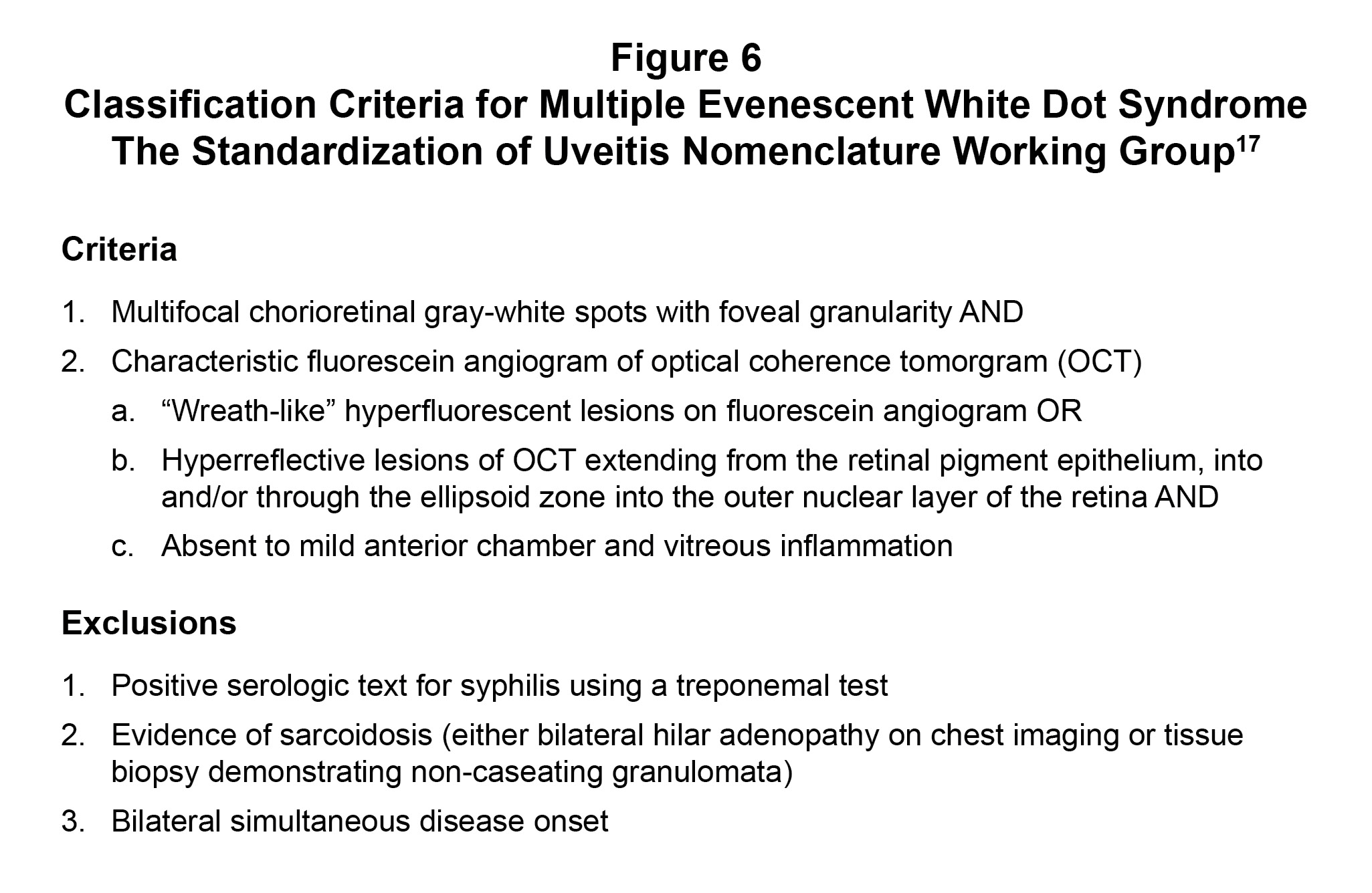
Figure 6. Classification Criteria for Multiple Evanescent White Dot Syndrome created by the Standardization of Uveitis Nomenclature Working Group. Click to enlarge
In 2021, The Standardization of Uveitis Nomenclature (SUN) Working Group identified classification criteria for multiple evanescent white dot syndrome based on machine learning of cases with MEWDS and 8 additional posterior uveitides.17 The goal of the SUN criteria is classification at presentation, so these criteria are most appropriate for the early stage of the condition. It is important to note that these classification criteria are different from clinical diagnostic criteria in that specificity is emphasized rather than sensitivity, which is more typically emphasized in diagnostic criteria. Figure 6 shows the classification criteria as published by the SUN Working Group.
The patient in this case meets both classification criteria. For criteria one, he had multifocal white spots in the right eye with foveal granularity seen on clinical exam and widefield color photography. For criteria two, he had hyperreflective lesions on OCT in the RPE that extended through to the outer nuclear layer as well as mild vitritis (b and c). While further testing was not done for syphilis or sarcoidosis, the patient met the exclusion criteria for lacking bilateral simultaneous disease onset.
In a 2020 retrospective, multicenter, case series on MEWDS masqueraders by Russel et al.,18 syphilitic outer retinitis was the most common masquerader. In a 2021 systematic review of the literature on syphilitic outer retinitis, the median age of presentation was 48 years.8 This is older than the typical patient demographics of MEWDS and much older than the teenage patient in this case. Over a third of patients in the review had visual acuity of 20/200 or worse, and all patients who presented with vitritis had acuity of 20/200 or worse.8 Both conditions can present with hyperautofluorescent lesions seen as white spots in the retina and ellipsoid zone disruption. However, patients with syphilitic outer retinitis tended to present bilaterally, rather than unilaterally, and resolve with penicillin treatment rather than spontaneously, as was seen in this case.18 Recommended testing for patients suspicious of syphilis infection includes a treponemal test, such as a fluorescent treponemal antibody absorption test (FTA-ABS) or the Treponema pallidum passive particle agglutination assay (TP-PA). Patients should also be tested for human immunodeficiency virus (HIV) due to the high rate of coinfection with syphilis. Syphilitic outer retinitis is considered a form of neurosyphilis and therefore should be treated with intravenous penicillin G for 2 weeks.8
Primary vitreoretinal lymphoma was the second most common masquerader in the same case series.18 However, these patients were also older and lacked spontaneous resolution seen in MEWDS. Fluorescein angiogram and fundus autofluorescence showed mottled hypo and hyperfluorescent spots in a “leopard spot” pattern, which is classic for lymphoma. Another masquerader of MEWDS was idiopathic multifocal choroiditis. Idiopathic multifocal choroiditis also affects young myopic women with reported symptoms of photopsias and blurry vision. This inflammatory choroiditis can leave behind RPE atrophy, pigmentary scars and lead to choroidal neovascular membranes. Compared to MEWDS, multifocal choroiditis is more commonly bilateral and associated with greater anterior chamber and vitreous cells.18
Management
MEWDS is a self-limiting condition that does not require medical therapy. Typically, visual symptoms and posterior segment changes on multimodal imaging resolve within 3 months.1,9, 14 It has therefore been described as the “common cold of the retina”.19
In a retrospective study of the clinical course of MEWDS and factors influencing visual recovery, reduced presenting visual acuity (VA) and younger age were associated with poorer final VA. The authors also concluded that the disruption of the outer retinal layers on OCT was not statistically significant in predicting the final VA as some patients had persistent disruption on OCT but complete visual recovery.20 Although the patient in this case was young, he presented with good visual acuity and continued to have good acuity as his lesions and symptoms resolved over the subsequent weeks.
Short pulse corticosteroids have been reported in a limited number of cases.21, 22 Patients have exhibited an improvement in visual acuity within 1 week and full visual recovery within 3 weeks.21 However, further prospective studies are needed to fully assess the clinical value of corticosteroids in MEWDS.
Conclusion
This teaching case report exemplifies how a seemingly routine appointment can result in an uncommon diagnosis. Clinicians should be aware of Multiple Evanescent White Dot Syndrome, its differential diagnoses and utilize available multimodal imaging to make the final diagnosis. Careful evaluation of multimodal imaging findings was key in distinguishing this diagnosis from others that can present similarly, especially when the patient is of an atypical demographic for all differential diagnoses. This teaching case report walks trainees and educators through the case with prompts for discussion of differential diagnoses, multimodal imaging interpretation, the final diagnosis and management of MEWDS based on the current literature.
Appendix
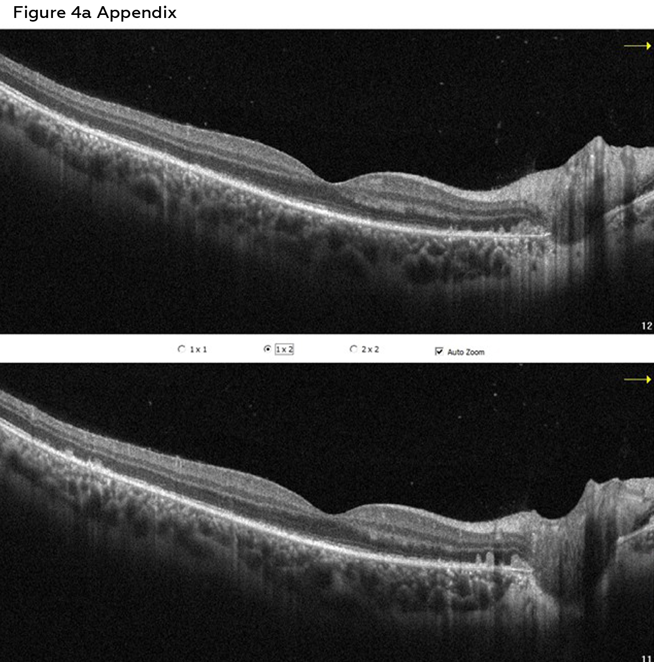
Appendix. Figure 4a Macula OCT right eye, visit two – without arrow annotations . Click to enlarge
References
- Jampol LM, Sieving PA, Pugh D, Fishman GA, Gilbert H. Multiple evanescent white dot syndrome. I. Clinical findings. Arch Ophthalmol. 1984 May;102(5):671-674. DOI: 10.1001/archopht.1984.01040030527008
- Pichi F, Srivastava S, Chexal S, et al. En face optical coherence tomography and optical coherence tomography angiography of multiple evanescent white dot syndrome: New Insights into Pathogenesis. Retina. 2016 Dec;36 Suppl 1:S178-S188. DOI: 10.1097/IAE.0000000000001255
- Papasavvas I, Mantovani A, Tugal-Tutkun I, Herbort CP Jr. Multiple evanescent white dot syndrome (MEWDS): update on practical appraisal, diagnosis and clinicopathology; a review and an alternative comprehensive perspective. J Ophthalmic Inflamm Infect. 2021 Dec 18;11(1):45. DOI: 10.1186/s12348-021-00279-7
- Herbort CP. Multiple evanescent white dot syndrome (MEWDS). In: Zierhut M, Pavesio C, Ohno S, Orefice F, Rao N, editors. Intraocular Inflammation. Berlin: Springer; 2016. pp. 997-1005. DOI: 10.1007/978-3-540-75387-2
- Sadda SR, Schachat AP, Wilkinson CP, et al. Editors. Ryan’s Retina. 7th ed. Philadelphia: Elsevier Health Sciences; 2022.
- Ahn SJ, Woo SJ, Kim KE, Jo DH, Ahn J, Park KH. Optical coherence tomography morphologic grading of macular commotio retinae and its association with anatomic and visual outcomes. Am J Ophthalmol. 2013 Nov;156(5):994-1001. DOI: 10.1016/j.ajo.2013.06.023
- Tang W, Guo J, Liu W, Xu G. Quantitative analysis of retinal and choriocapillary vascular density of multiple evanescent white dot syndrome by optical coherence tomography angiography. Graefes Arch Clin Exp Ophthalmol. 2020 Aug;258(8):1697-1707. DOI: 10.1007/s00417-020-04687-8
- Amaral C, Joy L, Jimenez H, Cruz-Inñigo YJ, Ulloa-Padilla JP, Oliver AL. Syphilitic outer retinopathy: a case report and review of the literature. J Vitreoretin Dis. 2021 Jun 21;6(1):63-70. DOI: 10.1177/24741264211018300
- Dell’Omo R, Mantovani A, Wong R, Konstantopoulou K, Kulwant S, Pavesio CE. Natural evolution of fundus autofluorescence findings in multiple evanescent white dot syndrome: a long-term follow-up. Retina. 2010 Oct;30(9):1479-1487. DOI: 10.1097/IAE.0b013e3181d50cd3
- Zou Y, Kamoi K, Zong Y, Zhang J, Yang M, Ohno-Matsui K. Ocular inflammation post-vaccination. Vaccines (Basel). 2023 Oct 23;11(10):1626. DOI: 10.3390/vaccines11101626
- Zeng Y, Du Z, Shao C, Zhao M. Comprehensive insights into COVID-19 vaccine-associated multiple evanescent white dot syndrome (MEWDS): a systematic analysis of reported cases. Hum Vaccin Immunother. 2024 Dec 31;20(1):2350812. DOI: 10.1080/21645515.2024.2350812
- Borruat FX, Herbort CP, Spertini F, Desarnaulds AB. HLA typing in patients with multiple evanescent white dot syndrome (MEWDS). Ocul Immunol Inflamm. 1998 Mar;6(1):39-41. DOI: 10.1076/ocii.6.1.39.8084
- Pellegrini M, Veronese C, Bernabei F, et al. Choroidal vascular changes in multiple evanescent white dot syndrome. Ocul Immunol Inflamm. 2021 Feb 17;29(2):340-345. DOI: 10.1080/09273948.2019.1678650
- Dell’Omo R, Pavesio CE. Multiple evanescent white dot syndrome (MEWDS). Int Ophthalmol Clin. 2012 Fall;52(4):221-232. DOI: 10.1097/IIO.0b013e31826a99c7
- Mantovani A, Invernizzi A, Staurenghi G, Herbort CP Jr. Multiple evanescent white dot syndrome: a multimodal imaging study of foveal granularity. Ocul Immunol Inflamm. 2019;27(1):141-147. DOI: 10.1080/09273948.2017.1353104
- Feigl B, Haas A, El-Shabrawi Y. Multifocal ERG in multiple evanescent white dot syndrome. Graefes Arch Clin Exp Ophthalmol. 2002 Aug;240(8):615-621. DOI: 10.1007/s00417-002-0478-7
- Standardization of Uveitis Nomenclature (SUN) Working Group. Classification criteria for multiple evanescent white dot syndrome. Am J Ophthalmol. 2021 Aug;228:198-204. DOI: 10.1016/j.ajo.2021.03.050
- Russell JF, Pichi F, Scott NL, et al. Masqueraders of multiple evanescent white dot syndrome (MEWDS). Int Ophthalmol. 2020 Mar;40(3):627-638. DOI: 10.1007/s10792-019-01223-4
- Tavallali A, Yannuzzi LA. MEWDS, common cold of the retina. J Ophthalmic Vis Res. 2017 Apr-Jun;12(2):132-134. DOI: 10.4103/jovr.jovr_241_16
- Bosello F, Westcott M, Casalino G, et al. Multiple evanescent white dot syndrome: clinical course and factors influencing visual acuity recovery. Br J Ophthalmol. 2022 Jan;106(1):121-127. DOI: 10.1136/bjophthalmol-2020-317357
- Norooznezhad AH, Mohammadzadeh V, Kadivar S, Ghassemi F. Multiple evanescent white dot syndrome: a case report and experience with corticosteroid therapy. Iran J Allergy Asthma Immunol. 2020 May 17;19(S1):91-94. DOI: 10.18502/ijaai.v19i(s1.r1).2865
- Takahashi Y, Ataka S, Wada S, Kohno T, Nomura Y, Shiraki K. A case of multiple evanescent white dot syndrome treated by steroid pulse therapy. Osaka City Med J. 2006 Dec;52(2):83-86.