Secondary Canaliculitis Due to Punctal Plugs
Alaina R. Bandstra, OD, FAAO
Secondary canaliculitis is a rare subcategory of canaliculitis, the inflammation of the proximal lacrimal pathway, commonly associated with punctal or intracanalicular plug use for dry eye management. These lacrimal plugs can elicit an infection within the canaliculus by damaging the mucosal tissue. In the presence of tear flow stasis, this enables bacterial proliferation. This teaching case report reviews the clinical presentation of canaliculitis, including signs that differentiate it from other anterior segment conditions. It also discusses how early diagnosis and appropriate medical management may prevent the need for surgical intervention.
Keywords
Background
Canaliculitis is a very rare inflammation of the lacrimal pathway in which an infection develops within the punctum and canaliculus, the two most proximal components of the nasolacrimal tear drainage system.1-5
Cases of canaliculitis are classified as either primary, with no known underlying etiology, or secondary, caused by lacrimal pathway obstruction.4,5 In most cases of secondary canaliculitis, lacrimal plugs are the inciting obstruction.3,5-8 Signs and symptoms associated with both primary and secondary canaliculitis are often mild and similar in presentation to those of other, more common, anterior segment diseases. These similarities frequently cause a delay in diagnosis or the misdiagnosis of canaliculitis, which can exacerbate the condition and result in poor outcomes.1-3,6 With prompt diagnosis, canaliculitis can be effectively treated with conservative management strategies; however, recurrence is common, and more invasive procedures, such as canaliculotomy, are often required for complete resolution.1,3,8-10 Accurate identification of the clinical features of canaliculitis, including the classic “pouting punctum”, aids in proper diagnosis, which may limit the need for surgical intervention.11
This teaching case report demonstrates the importance of early diagnosis and prompt treatment of secondary canaliculitis to avoid surgical intervention. It reviews the role that primary care optometrists play in managing this rare condition and is suitable for teaching or reviewing the condition with third- and fourth-year optometry students, optometry residents and novice practitioners with knowledge of the anatomical pathway of the nasolacrimal drainage system and an understanding of microbiological and pharmacological interactions.
Case Description
Initial Presentation
A 43-year-old Caucasian female presented with complaints of a red, tender and irritated lower right eyelid for the past month along the nasal lid margin near the medial canthus. She admitted to frequent lid rubbing, which exacerbated symptoms and often resulted in mild pain, tearing and burning of the affected eyelid. She denied discharge or vision changes. She denied any relief following consistent use of warm compresses, artificial tears and topical ocular allergy drops. Her ocular history included bilateral laser in situ keratomileusis (LASIK), intermittent alternating exotropia and dry eye syndrome. The dry eye was managed with artificial tears and the recent insertion of punctal plugs. Nine months prior to presentation, 0.6 mm petite silicone punctal plugs (brand name and manufacturer unspecified) were inserted into both lower puncta. At the time of presentation, the redness, tenderness and irritation symptoms were localized to the area around the right punctal plug. The patient’s medical history included depression, anxiety and migraines. She reported use of the following medications: divalproex sodium, rizatriptan, vortioxetine, trazodone, ketorolac and onabotulinumtoxinA injections. She noted an allergy to sulfa drugs. Her family history included diabetes (mother, grandfather) and glaucoma (grandmother).
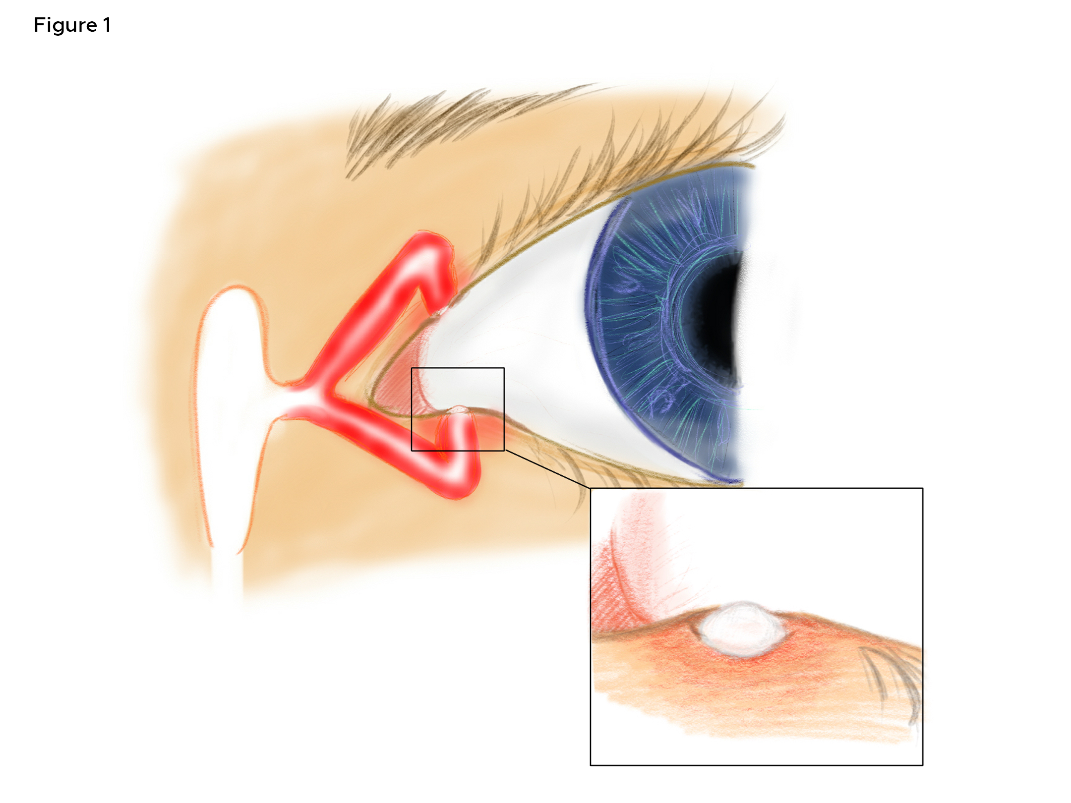
Figure 1 Diagram of anatomy of the puncta and lacrimal drainage, showing location of “pouting punctum” (inset) characteristic of canaliculitis. Click to enlarge
Entering uncorrected Snellen visual acuities were 20/25 in the right and left eyes, with pinhole acuities of 20/20 in each eye. Pupil, confrontation field and extraocular motility evaluations were unremarkable, and the intraocular pressure was 9 mmHg in each eye. Slit lamp evaluation revealed LASIK flap scarring and a low tear meniscus in each eye. The left lower punctal plug was absent. There was 1+ lid erythema and edema surrounding a punctal plug protruding from the right lower punctum, extending temporally towards the central lid. The inflammation was concentrated at the nasal lid margin and created the appearance of a swollen “pouting punctum” around the exposed plug (Figure 1).
Lid scaling, tenderness upon palpation and trace nasal conjunctival injection were present in the right eye. The posterior segment evaluation was unremarkable.
Given the localized inflammation surrounding the punctal plug in the right eye, removal of the plug was recommended to further investigate the etiology of the patient’s symptoms. Informed consent was obtained, and sterile jeweler’s forceps were used to extract the plug from the lower punctum without complication. Immediately following extraction, spontaneous excretion of purulent discharge from the punctum was observed. Gentle pressure to the lower eyelid aided in the complete expression of the discharge. No solid concretions, blood or foreign material was noted. Based on the observation of infectious inflammation (erythema, edema and purulent discharge) within the lacrimal pathway above the medial canthal tendon associated with a punctal plug, the patient was diagnosed with secondary canaliculitis of the right lower eyelid.
Treatment and Management
Treatment was initiated with a 1-week course of both oral cephalexin 500 mg taken twice daily and topical combination trimethoprim sulfate and polymyxin B sulfate ophthalmic solution for use in the right eye 4 times daily. The patient was also instructed to keep the eye clean by limiting makeup use and using lid scrubs 1-2 times daily. Future use of punctal plugs for dry eye management was discouraged. The patient continued dosing artificial tears up to 4 times daily in each eye. She was scheduled for a 5-day follow-up visit.
Follow-Up Care
Five days later, the patient reported compliance with all treatments as prescribed and denied any residual redness, tenderness, pain, discharge or swelling of the right lower eyelid. Her visual acuities were stable, and entrance testing was normal. Anterior segment evaluation revealed clear and open puncta in both eyes without evidence of inflammation of the right lower eyelid or canaliculus.
The patient was instructed to continue the oral and topical antibiotics for an additional 2 days to complete the full 1-week course. Given the near resolution of the canaliculitis, she was scheduled for follow-up in 2 months, with advice to return if symptoms worsened.
At the 2-month follow-up, the patient had completed and then discontinued all antibiotics (topical and oral) as instructed and reported no recurrence of symptoms. She did note persistent bilateral dry eye symptoms in the morning upon awakening, which resolved with the instillation of artificial tears. She declined further dry eye evaluation and was not interested in more aggressive management strategies at that time.
Anterior segment evaluation was unremarkable except for LASIK flap scarring, trace-1+ punctate keratitis and a reduced tear meniscus height in each eye. Both lower puncta were clear and open, and there was no expression with gentle pressure. Thus, the canaliculitis had fully resolved. The risk of recurrent canaliculitis was discussed, and the patient was instructed to return should she experience symptoms of tearing, discharge, redness or swelling of the eyelid. For management of her dry eye disease, she was encouraged to continue with regular dosing of artificial tears.
The patient returned for her annual exam 9 months later (1 year after initial onset) with a quiet anterior segment and no recurrence.
Education Guidelines
Key Concepts
- A complication of punctal plugs is secondary canaliculitis
- Consider canaliculitis as a differential diagnosis in chronic or recurrent medial eyelid lesions, conjunctivitis or blepharitis
- Identification of the classic, swollen “pouting punctum” helps differentiate canaliculitis from these conditions
- First-line medical management involves topical and oral antibiotics
- Appropriate diagnosis and early intervention may prevent the need for more invasive surgical intervention
Learning Objectives
At the conclusion of this case report, readers should be able to:
- Identify the clinical signs of canaliculitis, including the classic, swollen “pouting punctum”
- Understand how the pathophysiology of canaliculitis differs from Meibomian gland diseases, nasolacrimal duct obstruction and dacryocystitis
- Prescribe an appropriate first-line medical management protocol for canaliculitis
Discussion Questions
- Anatomy and pathophysiology of the nasolacrimal tear drainage system
- Describe the anatomy of the nasolacrimal tear drainage system, including the anatomical location of the puncta, canaliculi and lacrimal sac.
- What is the difference (in name) between an infection of the proximal aspect of this pathway vs. the distal aspect?
- How do infections of the various components of this system differ in their presentation?
- Clinical presentation of canaliculitis
- What is the difference between primary and secondary canaliculitis?
- What clinical symptoms and signs are typically present with this condition, and how is the diagnosis made?
- What pathogens commonly cause canaliculitis infections?
- Which anterior segment conditions should be considered in the differential diagnosis of canaliculitis, and how does a clinician differentiate between these diseases?
- Optometry’s role in the management of canaliculitis
- What is the first-line treatment for canaliculitis? Give specific examples of what may be prescribed.
- How does initial management differ in cases of secondary disease (compared to primary disease)?
- What is the primary risk associated with delayed diagnosis (and delayed treatment)?
Assessment of Learning Objectives
This teaching case report is intended for third- and fourth-year optometry students, optometry residents and novice practitioners with knowledge of the anatomical pathway of the nasolacrimal drainage system, microbiology and pharmacology. In an academic setting, it is recommended that a faculty member or residency supervisor facilitate the discussion in a small group, such as at a grand rounds presentation or clinical case discussion in the primary care, ocular disease or urgent care clinical setting. The case could also be presented in the classroom via a slide show presentation and facilitation of independent review or large-group discussion. The details of the case should be presented to learners with an opportunity to answer discussion questions prior to the formal case discussion. An open-ended discussion generates an environment where students and residents can gauge their knowledge gaps and learn from peers and instructors.
Discussion
Canaliculitis is a rare inflammatory disease of the proximal lacrimal pathway that accounts for only 2.0-4.0% of all lacrimal diseases and 0.8% of all lacrimal infections.1-5,12 The upper and lower canaliculi function as part of the tear drainage system within the lacrimal pathway.3,13 Each canaliculus originates at the punctal opening and transverses the medial lid margin vertically, then horizontally, for approximately 10.0 mm before converging into the common canaliculus. Tears flow from the common canaliculus through the lacrimal sac into the nasolacrimal duct before draining into the nasal cavity (Figure 2). An infection of the proximal portion of this pathway (the canaliculus and adjacent punctum) results in canaliculitis.
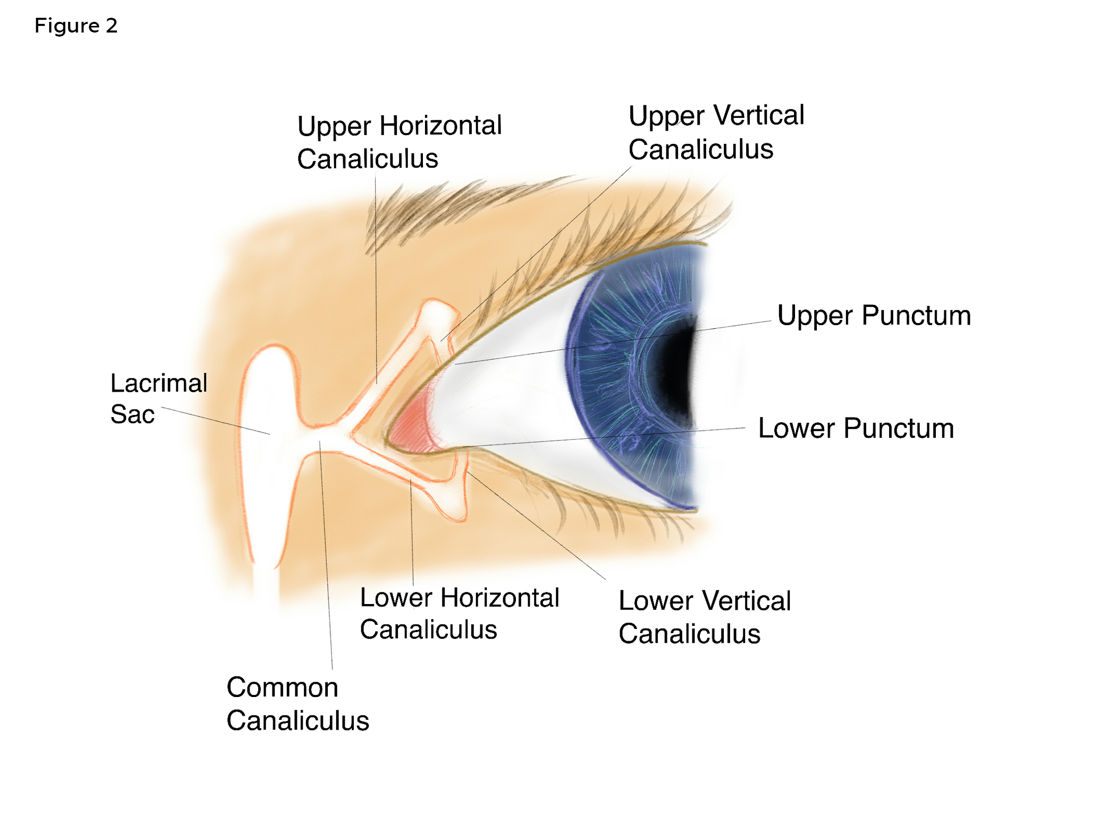
Figure 2. Diagram of the anatomy of the nasolacrimal tear drainage system. Click to enlarge
Due to the rare nature of canaliculitis, and its similar presentation to conjunctivitis, dacryocystitis and hordeolum, it is often misdiagnosed.1-3,6,12 A 2024 study reported that canaliculitis may initially be misdiagnosed in up to 60% of cases.12 A similar study conducted in 2011 found that up to 33% of cases were misdiagnosed multiple times.3 Delayed diagnosis has been reported up to 20 years after the onset of symptoms3 and commonly spans more than 5 months.5,6,9,10 This often results in recommendation of inappropriate treatments, which may further exacerbate the disease process.2,3
Presenting symptoms of canaliculitis most commonly include epiphora and discharge, while ocular irritation or mild pain, conjunctival injection and swelling, and erythema of the medial lid margin are also possible.1-5,9,10,12 Symptoms are typically unilateral and associated with a purulent or mucopurulent discharge. Purulence refers to the presence of white blood cells (pus) within a secretion, which indicates an infectious etiology. When secreted from mucous membranes (such as the lacrimal pathway), the term mucopurulent may be used to describe the addition of mucus to this discharge. A classic “pouting punctum” is considered pathognomonic.5,12 In addition to generalized inflammation of the nasal lid margin and adjacent conjunctiva, punctal discharge may include yellow, gritty sulfur granules, known as concretions, which are diagnostic of canaliculitis (though present in only one-third of cases).1-3,7,9-12 Given the high rate of misdiagnosis, Singh et al proposed the use of a “clinical tetrad” to aid in the diagnosis of lacrimal canaliculitis, which emphasizes identification of the clinical signs of canaliculitis, including medial eyelid inflammation with a “pouting punctum” and punctal discharge.14
Infection occurs most commonly in the lower canaliculus (55-87%)4-6,10,12 and shows no right or left eye predilection.6,10,12 Canaliculitis typically presents in females 50-65 years of age;9,10,12 however, larger age ranges and pediatric cases have been reported.6,12 The strong female predilection (two to five times more common) is attributed to menopause-related hormonal changes, resulting in poor tear production with reduced antimicrobial properties.2,6,12 Further, frequent use of makeup and lacrimal plugs in this population enhances bacterial proliferation within the lacrimal pathway.
There are two forms of canaliculitis: primary and secondary. In primary canaliculitis, the infection occurs without foreign obstruction.4,5,12 It has been proposed that a diverticulum within the proximal lacrimal pathway obstructs tear drainage, allowing bacteria to accumulate; however, known predisposing factors are often difficult to identify. In secondary canaliculitis, a foreign body within the lacrimal drainage system initiates the infectious process.3-8,12 Injury to the canaliculus mucosa, in addition to tear flow stasis, creates a reservoir for bacterial growth. Lacrimal plugs (either punctal or intracanalicular) are the most common cause of secondary canaliculitis. In a literature review of canaliculitis, Pal et al. classified 91.59% of the reviewed cases as primary, noting SmartPLUG™ (Medennium Inc., Irvine, California; a thermosensitive material that allows the plug to expand and fit the canaliculus) use in 86.72% of secondary cases.12 In a similar review, Huang et al. found that cases of secondary canaliculitis had an even stronger female predilection than that of primary disease (100% vs. 65%).7
Secondary canaliculitis is more common with intracanalicular plugs; however, punctal plug-related cases have also been reported. In 2015, the American Academy of Ophthalmology reviewed the safety of lacrimal plugs for dry eye. In this report, the incidence of secondary canaliculitis was low in both groups (7.8% with intracanalicular plugs; 0.0-5.6% with punctal plugs).8 Huang et al. also found a higher incidence of canaliculitis in the intracanalicular plug group.7 This retrospective review of secondary canaliculitis reported that only 18% of cases were related to punctal plugs, while 82% were intracanalicular. The increased risk of inflammation associated with intracanalicular plugs is attributed to a more distal location within the lacrimal pathway, which more easily damages the mucosal tissue, allowing for bacterial proliferation. Regardless of plug type, continued monitoring is important, as most cases of secondary canaliculitis develop between 3 to 5.5 years after plug insertion.7
Diagnosis of canaliculitis is typically made based on clinical presentation alone.2,3 Given the frequency of misdiagnosis, canaliculitis should be considered in any patient presenting with chronic or recurrent medial eyelid inflammation, conjunctivitis or blepharitis, especially if the patient has a history of lacrimal plugs. In this case, the patient presented with symptoms consistent with canaliculitis; however, these symptoms are also common to other anterior segment diseases. A careful assessment of the clinical findings was necessary for proper diagnosis. The primary identifying feature was a classic “pouting punctum” of the right lower eyelid with surrounding lid inflammation. Removal of the punctal plug allowed expression of a purulent discharge from the punctal opening. Though concretions were not evident in the expressed material, the localized inflammation, presence of punctal discharge, and history of lacrimal plug use, along with the mild nature of the patient’s symptoms, supported the diagnosis of secondary canaliculitis.
Other conditions considered in this case include chalazion or hordeolum, acquired nasolacrimal duct obstruction (NLDO) and dacryocystitis.2,3,6,12,15 Meibomian gland obstruction (not lacrimal pathway obstruction) can cause chalazia or hordeola. Localized inflammation surrounding a palpable eyelid nodule is often evident; however, punctal discharge is never present. Acquired NLDO and dacryocystitis are important differentials in this case as they represent diseases of the lacrimal pathway; however, both differ in presentation from that of canaliculitis. An acquired NLDO refers to an obstruction of the tear drainage system that develops during adulthood. It presents with classic symptoms of persistent unilateral epiphora, without significant infection or inflammation. Dacryocystitis is an infection of the lacrimal sac. Symptoms of dacryocystitis include severe pain, erythema and edema of the distal portion of the lacrimal pathway and may be accompanied by a fever. In this case, the location of inflammation within the proximal lacrimal pathway and the mild nature of the patient’s symptoms excluded dacryocystitis (Figure 3).
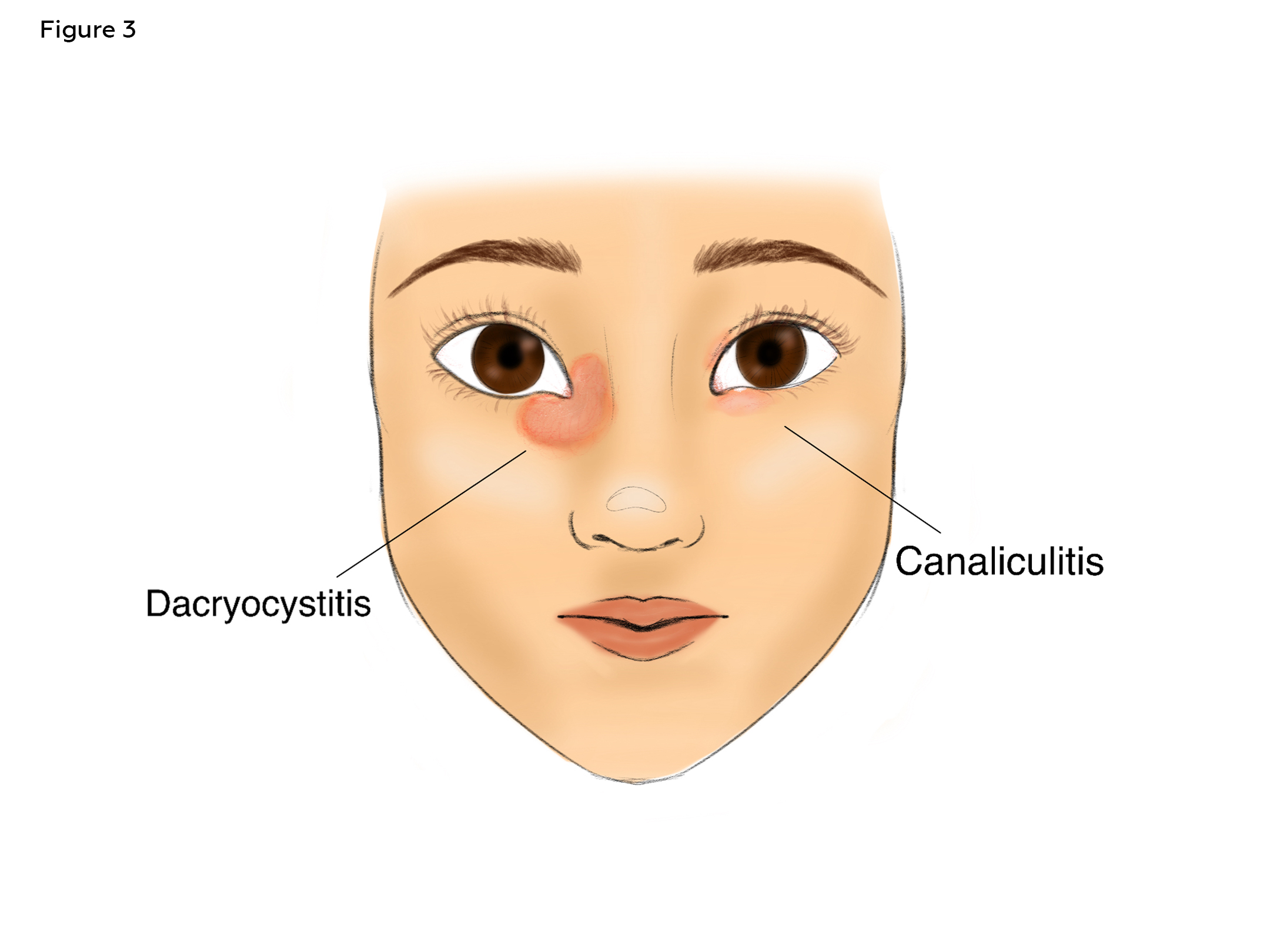
Figure 3. Diagram of the difference in clinical presentation between dacryocystitis (left) and canaliculitis (right). Click to enlarge
Initial medical management of canaliculitis is conservative with use of topical and oral antibiotics supported by warm compresses and local massage.1-3,12 In a retrospective review, Anand et al. reported a high susceptibility of all microorganisms isolated from canaliculitis cultures to gentamicin, chloramphenicol and cefotaxime.16 In a review by Huang et al., Streptococcus sp, P. acnes, and Staphylococcus sp (Gram-positive) were the most common isolates from primary canaliculitis, while Pseudomonas aeruginosa (Gram-negative) was most common in secondary disease, though mixed infections were frequent in all cases.7 Based on this study, prescribed antibiotics should provide coverage against both Gram-positive and Gram-negative organisms (particularly Pseudomonas aeruginosa). Well-tolerated oral antibiotics commonly used in the treatment of anterior segment diseases include doxycycline 100 mg taken twice daily for 7-14 days or cephalexin 500 mg taken twice daily for 7-14 days.17 Topical ophthalmic antibiotics to consider are gentamicin 0.3% (for its protection against Pseudomonas aeruginosa) taken 4 times daily for 7-14 days or combination trimethoprim sulfate and polymyxin B sulfate (a broad-spectrum agent) taken 4 times daily for 7-14days. Some reports also propose the addition of topical corticosteroids.18
In cases of primary canaliculitis, lacrimal duct irrigation with antibiotics or intracanalicular ophthalmic corticosteroid/antibiotic ointment infiltration (IOI) may also be considered.12 Caution should be exhibited when using lacrimal irrigation in suspected cases of secondary canaliculitis to avoid distal plug migration.2,3 For secondary canaliculitis, removal of the lacrimal plug is necessary prior to treatment.1-3,12 Punctal plugs are easily removed with forceps.19 Intracanalicular plugs, however, prove more challenging as they are most often removed via irrigation,19-21 which may force the plug to lodge more distally within the lacrimal pathway, increasing complications.2,3 Imaging is commonly performed prior to surgical intervention in cases of secondary canaliculitis in order to confirm the location of a retained intracanalicular plug.
Disease recurrence following conservative management of canaliculitis occurs in 33% of cases.1-3 Initiating treatment early improves success rates, but frequent delays in diagnosis often prevent prompt intervention.3 Higher rates of recurrence have also been linked to the presence of concretions,1-3,7,9,10,12 which obstruct topical antibiotic penetration and act as a reservoir for bacterial growth. Successful resolution of canaliculitis depends on complete concretion extraction; therefore, manual expression of punctal discharge should be attempted prior to initiating medical therapy.3
In this case, the patient was diagnosed with canaliculitis secondary to punctal plug insertion. Following plug removal and gentle expression of punctal discharge, oral cephalexin and topical combination trimethoprim sulfate and polymyxin B sulfate ophthalmic solution were prescribed. Cephalexin is a well-tolerated cephalosporin medication with broad-spectrum coverage and is frequently used for skin and soft tissue infections.17 Cephalosporins have been shown to be highly effective against most microorganisms isolated in canaliculitis cultures.16 Topical polymyxin has also been shown to successfully treat cases of primary and secondary canaliculitis,1 and is effective against Pseudomonas aeruginosa. Furthermore, when combined with trimethoprim sulfate, broad-spectrum topical coverage is achieved.17 In this case, the patient’s infection cleared rapidly within 5 days of initiating treatment, and there was no evidence of disease recurrence over the following year. The patient did not require more invasive surgical intervention.
Surgical intervention is indicated when conservative treatment fails or in cases of recurrent disease.12 Alternative treatments for primary canaliculitis include one-snip punctoplasty with curettage, lacrimal endoscopy, lacrimal duct irrigation with antibiotics, or intracanalicular ophthalmic corticosteroid/antibiotic combination infiltration (IOI).2,12,18,22 Canaliculotomy is the definitive treatment for canaliculitis.1-3,8,10 In secondary canaliculitis, a canaliculotomy also includes intracanalicular plug removal.7 Dacryocystorhinostomy (DCR) with silicone intubation may be required in more complicated cases, or if a plug has been forced distally due to lacrimal irrigation.7,20
The success of conservative therapy in this case is attributed to an early diagnosis with prompt intervention. In contrast to most cases of canaliculitis, treatment was initiated early in this case (within 1 month of symptom onset). Furthermore, there was no evidence of concretions in the expressed punctal discharge, which are known to harbor bacteria and prolong the infection. This lack of concretions, in addition to the successful removal of the punctal plug early in the disease course, allowed the prescribed antibiotics to successfully penetrate the infected mucosa without the need for more invasive and high-risk surgical procedures.
Conclusion
Canaliculitis is a relatively uncommon condition, but it can be successfully triaged and managed by primary eyecare providers from onset. Understanding the presenting clinical features of canaliculitis and differentiating it from other anterior segment disorders is crucial to the successful management of this lacrimal pathway disease. Avoid any delay in diagnosis by recognizing characteristic signs of canaliculitis, which include the classic, swollen “pouting punctum.” Early intervention and medical management improve successful resolution and can help avoid the need for more invasive procedures. Secondary canaliculitis is often caused by lacrimal plugs. Extraction of the plug, followed by complete expression of any purulent material in the punctum, will improve antibiotic access to the infected tissues, thus quickly and successfully eliminating the canaliculitis without the need for more invasive surgical intervention.
Acknowledgements
The author would like to acknowledge Lindsay A. Sicks, OD, FAAO, FIACLE, FSLS, for editing assistance and Vivian La for the original illustrations.
References
- Zaveri J, Cohen AJ. Lacrimal canaliculitis. Saudi J Ophthalmol. 2014 Jan;28(1):3–5. DOI: 10.1016/j.sjopt.2013.11.003
- Kim UR, Wadwekar B, Prajna L. Primary canaliculitis: The incidence, clinical features, outcome and long-term epiphora after snip-punctoplasty and curettage. Saudi J Ophthalmol. 2015 Oct-Dec;29(4):274–7. DOI: 10.1016/j.sjopt.2015.08.004
- Freedman JR, Markert MS, Cohen AJ. Primary and secondary lacrimal canaliculitis: a review of literature. Surv Ophthalmol. 2011 Jul;56(4):336–47. DOI: 10.1016/j.survophthal.2010.12.001
- Balıkoğlu Yılmaz M, Şen E, Evren E, Elgin U, Yılmazbaş P. Canaliculitis awareness. Turk J Ophthalmol. 2016 Jan;46(1):25–9. DOI: 10.4274/tjo.68916
- Alam MS, Poonam NS, Mukherjee B. Outcomes of canaliculotomy in recalcitrant canaliculitis. Saudi J Ophthalmol. 2019 Jan-Mar;33(1):46–51. DOI: 10.1016/j.sjopt.2018.12.001
- Gogandy M, Al-Sheikh O, Chaudhry I. Clinical features and bacteriology of lacrimal canaliculitis in patients presenting to a tertiary eye care center in the Middle East. Saudi J Ophthalmol. 2014 Jan;28(1):31–5. DOI: 10.1016/j.sjopt.2013.09.006
- Huang YY, Yu WK, Tsai CC, Kao SC, Kau HC, Liu CJL. Clinical features, microbiological profiles and treatment outcome of lacrimal plug-related canaliculitis compared with those of primary canaliculitis. Br J Ophthalmol. 2016 Sep;100(9):1285–9. DOI: 10.1136/bjophthalmol-2015-307500
- Marcet MM, Shtein RM, Bradley EA, et al. Safety and efficacy of lacrimal drainage system plugs for dry eye syndrome: A report by the American academy of ophthalmology. Ophthalmology. 2015 Aug;122(8):1681–7. DOI: 10.1016/j.ophtha.2015.04.034
- Zhang Q, Xu B, Li XX, Li MW. Clinical characteristics, treatment patterns, and outcomes of primary canaliculitis among patients in Beijing, China. Biomed Res Int. 2015 Feb 17;2015:904756. DOI: 10.1155/2015/904756
- Xiang S, Lin B, Pan Q, et al. Clinical features and surgical outcomes of primary canaliculitis with concretions. Medicine (Baltimore). 2017 Mar;96(9):e6188. DOI: 10.1097/MD.0000000000006188
- Perumal B, Carlson JA, Meyer DR. A pathological analysis of canaliculitis concretions: More than just Actinomyces. Scientifica (Cairo). 2016 Jun 14;2016:6313070. DOI: 10.1155/2016/6313070
- Pal SS, Alam MS. Lacrimal canaliculitis: A major review. Semin Ophthalmol. 2024 Nov;39(8):586–94. DOI: 10.1080/08820538.2024.2354689
- Benitez-Del-Castillo JM, Lemp MA. Ocular Surface Disorders. London: JP Medical; 2013.
- Singh M, Mehta A, Sharma M, Kaur M, Gupta P. A “clinical tetrad” for easy diagnosis of lacrimal canaliculitis. J Curr Ophthalmol. 2022 Jul-Sep;34(3):347–51. DOI: 10.4103/joco.joco_307_21
- Gerstenblith AT, Rabinowitz MP. The Wills Eye Manual: Office and Emergency Room Diagnosis and Treatment of Eye Disease. 6th ed. Philadelphia: Lippincott Williams and Wilkins; 2012.
- Anand AR, Harinee R, Jeyalatha MV, et al. Microbiological profile of canaliculitis and their antibiotic susceptibility patterns: A 11-year review at a referral eye care centre. Indian J Med Microbiol. 2022 Jul-Sep;40(3):378–83. DOI: 10.1016/j.ijmmb.2022.05.007
- Bartlett JD, Jaanus SD. Clinical Ocular Pharmacology. 5th ed. St Louis, MO: Elsevier; 2008.
- Xu J, Liu Z, Mashaghi A, et al. Novel therapy for primary canaliculitis: A pilot study of intracanalicular ophthalmic corticosteroid/antibiotic combination ointment infiltration. Medicine (Baltimore). 2015 Sep;94(39):e1611. DOI: 10.1097/MD.0000000000001611
- Jehangir N, Bever G, Mahmood SMJ, Moshirfar M. Comprehensive Review of the Literature on Existing Punctal Plugs for the Management of Dry Eye Disease. J Ophthalmol. 2016;2016:1–22. DOI: 10.1155/2016/9312340
- Bourkiza R, Lee V. A review of the complications of lacrimal occlusion with punctal and canalicular plugs. Orbit. 2012 Apr;31(2):86–93. DOI: 10.3109/01676830.2011.648802
- Klein-Theyer A, Boldin I, Rabensteiner DF, Aminfar H, Horwath-Winter J. Prevalence of canaliculitis after smartplug insertion during long-term follow-up. Br J Ophthalmol. 2015 Aug;99(8):1134–6. DOI: 10.1136/bjophthalmol-2014-306290
- Zheng Q, Shen T, Luo H, et al. Application of lacrimal endoscopy in the diagnosis and treatment of primary canaliculitis: Practical technique and graphic presentation: Practical technique and graphic presentation. Medicine (Baltimore). 2019 Aug;98(33):e16789. DOI: 10.1097/MD.0000000000016789